
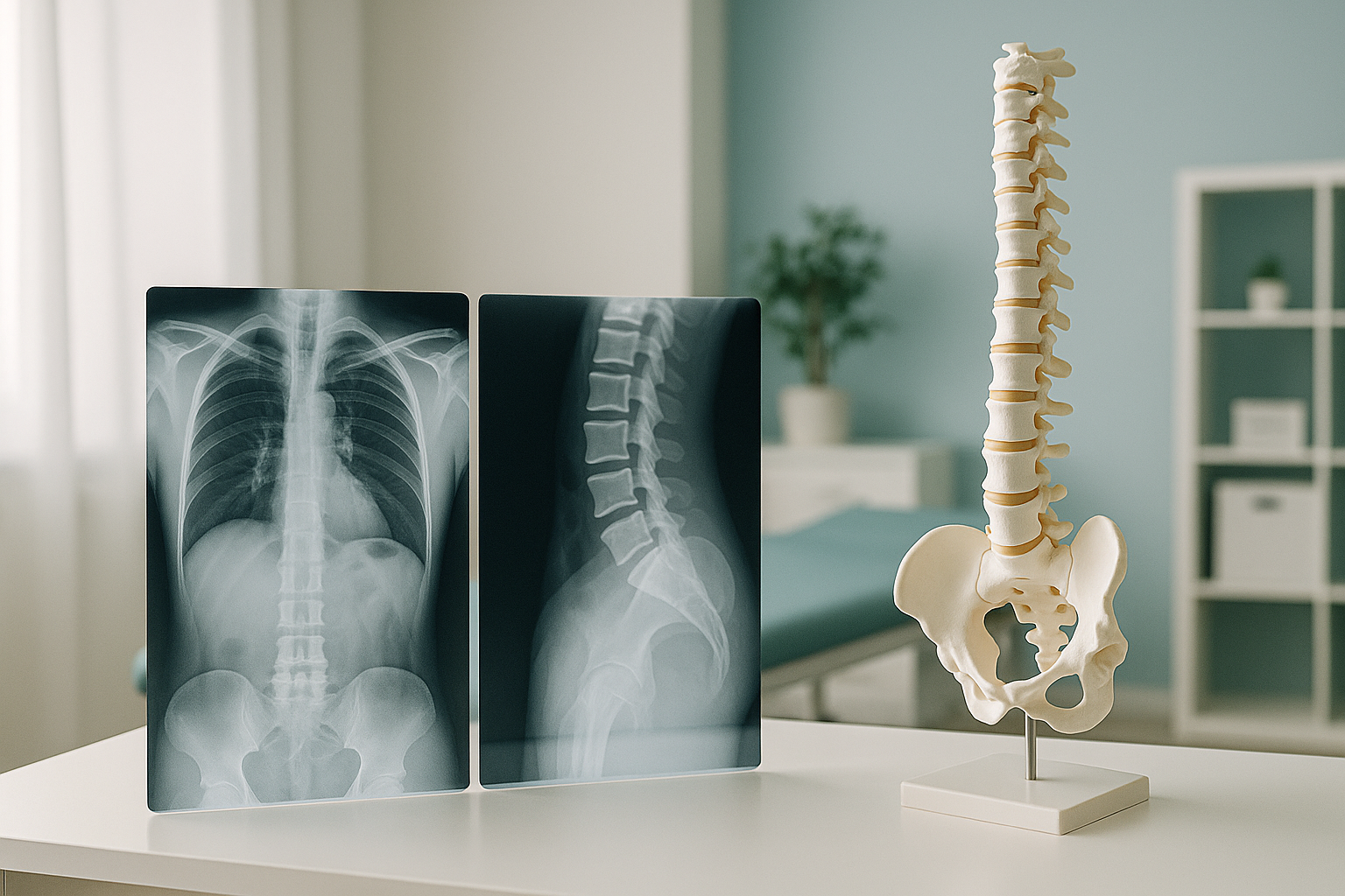
How Do Two X-rays Become a 3D Model of Your Skeleton?
An EOS scanner takes a front view and a side view of your body at the same time while you stand. Special software joins these two flat images into a detailed 3D model of your spine and a full rebuild of your skeleton. This lets your clinician see curves, rotation and alignment that a single flat X-ray cannot show.
Key points
- The scanner needs only two images taken at the same time, one from the front and one from the side.
- Software maps your bones onto a calibrated 3D model of the skeleton that can be turned and examined.
- You stand naturally during the scan, so the model shows your real alignment while your body carries its weight.
- The process uses about 90% less radiation than a standard X-ray, around ten times less.
- No extra dose is needed. The 3D model comes from the same two images.
How does biplanar imaging create a 3D reconstruction?
Two X-ray sources capture your whole body from two directions at the same moment. This method is called biplanar imaging. The scanner's geometry is precisely calibrated, so the software knows exactly how the front and side views relate to each other. It uses this to work out where each bone sits in three-dimensional space. It then wraps a surface model around the data. The result is a 3D model of your skeleton that you and your clinician can turn on screen. If this type of scan is new to you, read more about what an EOS scan is.
The four steps
You step into the scanner
You stand in your normal posture inside an open booth. There is no tunnel and nothing touches you.
Two beams move from head to toe
A front beam and a side beam move together and capture your whole skeleton in one pass. The scan takes roughly 15 to 20 seconds.
Software builds the 3D model
Using the calibrated geometry of the two views, the software works out every vertebra, the pelvis, the hips and the legs in three dimensions. No extra radiation dose is needed.
Your clinician reviews the model
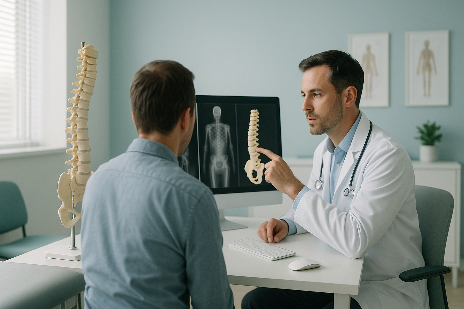
The 3D model can be turned and examined from any angle. Learn more about how EOS scan results are reported.
What can a 3D spine model show that a flat X-ray cannot?
A standard X-ray gives one flat view. You can see whether the spine curves to the left or right. But you cannot tell how much it twists, or how the pelvis tilts forward or backward. A 3D model of the spine adds depth to the picture. This lets your clinician assess rotation, torsion and pelvic measurements all at once.
How severe the spinal curve is
Precise Cobb angle measurement while your body carries its weight.
Rotation of the vertebrae
Twist that cannot be seen on a single flat image can be measured in the 3D model.
Pelvic tilt and balance
Shows how your pelvis sits in relation to your hips, spine and centre of gravity.
Leg length and alignment
The full-body image captures both legs so they can be compared directly, side by side.
These measurements are especially useful for understanding anteversion versus retroversion in the hip. They also help track a spinal curve over time without adding to radiation exposure.
When MRI is the better choice: EOS and its 3D model show bones, joints and alignment in excellent detail. For soft-tissue problems such as disc bulges, nerve compression or ligament tears, MRI is usually the better tool. The two scans answer different questions and often work well together.
How does a 3D standing scan compare with other imaging?
The biggest difference is that an EOS scan captures your skeleton while you stand. Imaging done lying down, such as CT or MRI, relaxes your muscles and lets your joints shift. Alignment problems that only appear under your full body weight can be missed completely.
| Feature | Standard X-ray | CT scan | EOS with 3D |
|---|---|---|---|
| Position | Standing (one view) | Lying down | Standing (two views at the same time) |
| 3D model | No | Yes | Yes |
| Full body in one image | Rarely | No (in slices) | Yes |
| Alignment under body weight | Partial | No | Yes |
| Radiation | Standard dose | Higher dose | About 90% less than standard X-ray |
The body is a connected chain from the feet to the head. So a problem in one place often makes another area compensate. A full standing image with a 3D model lets your clinician see the whole chain at once, rather than guessing from one area scanned on its own.
If you are about to start treatment, or are thinking of changing the one you are on, it is worth getting a detailed diagnosis first. Knowing whether the cause sits in the pelvis, the legs or the spine changes what will actually help.
Would you like to see your alignment in 3D? Book a 3D standing scan or start with a free video consultation to find out whether it is right for you.
Book a free video consultationWhat is included in a 3D standing scan at ScanAlign?
Every scan at ScanAlign standing EOS imaging includes the full 3D model as standard. There is no extra charge and no extra radiation dose for the 3D model.
- Full-body or targeted biplanar imaging while you stand.
- 3D model of the spine and the legs from the same images.
- A detailed report by a specialist radiographer, covering alignment, curves and key measurements.
- Images and report shared digitally, so you and your treating clinician both have a copy.
The scan takes place at 19 Harley Street, London. You do not need a referral from your doctor. You can book directly yourself.
Frequently asked questions
The scanner takes a front image and a side image at the same moment. The scanner's geometry is precisely calibrated, so software can work out the three-dimensional position of every bone and wrap a surface model around the data.
Not exactly. A CT scan images the body in slices while you lie down, and uses a much higher radiation dose. The EOS 3D model is built from just two standing images, with about 90% less radiation than a standard X-ray, around ten times less. It shows alignment while your body carries its real weight, which CT cannot.
No. The 3D model is made entirely from the same two images taken during the scan. There is no additional exposure.
Biplanar imaging means taking two X-ray views, front-to-back and from the side, at exactly the same time. Taking them together is what makes the 3D calculation possible. It also makes sure both views show the same posture.
When you lie down, gravity no longer pulls on your spine and pelvis in the usual way. Muscles relax and joints shift. Standing imaging captures your skeleton as it actually behaves in daily life, so the 3D model gives a more accurate picture of your alignment.
No. The EOS 3D model focuses on bones and joints. For soft-tissue problems such as discs, nerves or ligaments, MRI is the better tool. The two types of scan often work well together.
About 90% less than a standard X-ray, around ten times less. This makes it one of the lowest-dose options available for imaging the skeleton.
No. ScanAlign accepts patients who book for themselves. You can book directly without seeing your doctor first.
The price for patients travelling from abroad is confirmed during the free video consultation. In that call you can also discuss whether the scan is suitable for you before committing.
A specialist radiographer reviews your images and 3D model and writes a detailed report. Both the images and the report are shared with you digitally, so you can pass them on to your treating clinician.
See your alignment in 3D
A free video consultation lets you ask questions and find out whether a standing 3D scan is right for you. No pressure, no obligation.
Book a free video consultationWritten by Abbas Dhami (Specialist Diagnostic Radiographer)
Sources
- Dubousset J, Charpak G, Dorion I, et al. A new 2D and 3D imaging approach to musculoskeletal physiology and pathology with low-dose radiation and the standing position: the EOS system. Bull Acad Natl Med. 2005;189(2):287-300.
- Illés T, Somoskeöy S. The EOS imaging system and its uses in daily orthopaedic practice. Int Orthop. 2012;36(7):1405-1411.
- European Society of Radiology (ESR). Medical imaging dose optimisation strategies. Insights Imaging. 2021.
This page is general information and not a diagnosis.
ScanAlign is the trading name of CoreMed Solutions Ltd and operates under The Harley Street Hospital's CQC licence.