

Cobb Angle Measurement: How Are Spinal Curves Assessed?
A Cobb angle measurement is the standard way doctors and radiographers check how much the spine curves to the side or from front to back. It is worked out from an X-ray or a standing scan. Lines are drawn along the most tilted vertebrae at the top and bottom of a curve. The angle this gives tells your clinician whether a curve is mild, moderate or significant.
Key Points
- The Cobb angle is the standard used worldwide to measure how the spine curves on imaging.
- It is used for sideways curves (scoliosis) and for front-to-back curves (kyphosis and lordosis).
- Measurements taken while you stand under load show real life better than those taken lying down.
- EOS whole-body standing imaging uses about 90% less radiation than a standard X-ray, around ten times less.
- A Cobb measurement alone does not explain the cause of pain. Whole-body alignment gives the bigger picture.
What is a Cobb angle?
A Cobb angle is a number that shows how much your spine curves in a certain direction. The orthopaedic surgeon John Robert Cobb developed it in 1948. It has been the standard way to measure spinal curves ever since. Your clinician may be looking at a sideways curve (scoliosis) or at too much forward or backward curve (kyphosis or lordosis). Either way, they will almost always report it as a Cobb angle.
Put simply, a larger number means a bigger curve. Your specialist uses this number to decide whether a curve needs watching, more tests, or advice from a spine team. It is not a diagnosis on its own. But it is a vital piece of the puzzle.

How is a Cobb angle measured?
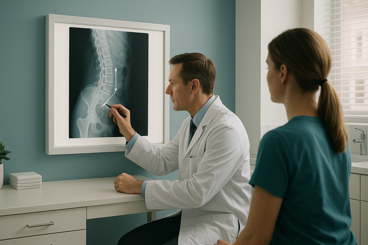
A radiographer or specialist finds the vertebrae at the very top and very bottom of the curve on an X-ray or standing scan. One line is drawn along the upper surface (endplate) of the top vertebra. Another is drawn along the lower surface of the bottom vertebra. The angle where these two lines meet is the Cobb angle.
This sounds technical, but the result is simple. Your report will describe the curve as mild, moderate or significant. Your clinician then looks at this together with your symptoms, posture and medical history to plan the next steps.
Often watched without treatment. Your clinician may suggest imaging from time to time to check for any change.
May call for physiotherapy, a specialist review or more detailed imaging to understand the cause and effect of the curve.
Usually needs a specialist spine assessment. The curve may be affecting breathing or balance, or causing persistent pain.
What signs suggest you might have a spinal curve?
Many spinal curves develop slowly. You may not notice one until someone else points it out, or until you start to feel its effects. Common signs include one shoulder higher than the other, uneven hips, clothes hanging unevenly, back pain or stiffness that does not get better with usual treatment, and a visible difference between the two sides when you bend forward.
Pain from a spinal curve does not always show up at the curve itself. Your body is a connected chain from feet to head. So a curve in the mid-back can make the pelvis tilt, and this puts extra load on one hip or knee. This is why a whole-body assessment is so valuable. It shows the ways your body has adjusted to make up for the curve.
Wondering whether your symptoms are linked to spinal alignment? A standing whole-body scan can give you a clear answer.
Book a free video consultationWhy does it matter whether a Cobb angle is measured standing or lying down?
It matters because gravity changes your spine. When you lie down for a usual X-ray, CT or MRI, your muscles relax. Your spinal curves can flatten or shift. So the measurement may not show how your spine really behaves during the day, when you are upright and carrying your weight.
A standing scan captures your spine under load, just as it works in real life. Research consistently shows that Cobb angles measured standing can differ meaningfully from those measured lying down. For your clinician, that difference can change the advice on what to do next.
How does EOS imaging help with Cobb angle measurement?
EOS is a standing, whole-body X-ray system. It captures your full skeleton in one pass, from head to feet, while you stand upright. This means the Cobb angle it measures shows your true spinal alignment under gravity. It also shows the rest of your skeleton at the same time. So your clinician can see how a spinal curve affects your pelvis, hips, knees and overall balance.
EOS uses about 90% less radiation than a standard X-ray, around ten times less. This makes it especially suitable if you need follow-up scans to watch a curve over time.
Because EOS images the whole body in one scan, it shows how your body has adjusted to a curve. A tilted pelvis, an uneven load on the legs, a shifted centre of gravity. These details cannot be seen on scans of a single area, but they can be the key to understanding why you have pain.
Before you start treatment, or change the one you are on, it is worth getting a detailed diagnosis. Knowing whether the cause sits in your pelvis, your legs or your spine changes what will actually help.
When MRI is the better choice: EOS shows bones, joints and alignment clearly. For soft-tissue problems such as slipped discs, pressure on nerves or ligament damage, MRI is usually the better tool. Your clinician can advise which type of imaging you need.
Want to understand your spinal curve as part of your whole-body alignment? Speak to our team first, free of charge, before you plan your trip to London.
Book a free video consultationLearn more about how EOS whole-body standing imaging works and what a scan involves.
Common questions about Cobb angle measurement
It is the standard way to measure how much the spine curves. A specialist draws lines on an X-ray or standing scan along the most tilted vertebrae at each end of the curve. The angle between those lines is the Cobb angle. A bigger number means a bigger curve.
No. It is also used to measure kyphosis (an excessive forward curve in the upper back) and lordosis (an excessive inward curve in the lower back). Any curve of the spine can be described with a Cobb angle.
Yes. When you lie down, gravity no longer presses down on your spine, and your muscles relax. This can make a curve look smaller than it really is. A standing measurement generally shows your day-to-day alignment better.
The radiographer finds the vertebra at the top of the curve that tilts most into the curve, and the one at the bottom. A line is drawn along the upper endplate of the top vertebra and the lower endplate of the bottom one. The angle where perpendicular lines from each cross is the Cobb angle.
No. You can book directly yourself. We first offer a free video consultation, so our specialist radiographer can confirm the scan is right for you and answer any questions. You do not need a referral from your doctor.
EOS uses about 90% less radiation than a standard X-ray, around ten times less. This makes it one of the lowest-dose imaging options available. You can find more detail on our FAQs and safety page.
ScanAlign is a private, self-pay clinic. The price for patients travelling from abroad is confirmed during the free video consultation, and you can read more on our FAQs page.
On its own, no. A Cobb measurement tells you the size of a curve, not the cause of your pain. That is why whole-body alignment imaging is so useful. It shows how a spinal curve is affecting the rest of your body. It reveals how the pelvis, hips and knees have adjusted, and these changes may be driving your symptoms.
A larger curve usually means a specialist review is a good idea. ScanAlign provides the diagnostic imaging and a detailed report. Your clinician, whether an orthopaedic surgeon, a physiotherapist or your family doctor, then uses that information to guide any treatment decisions.
Yes. The low radiation dose makes EOS especially suitable for follow-up imaging. Repeat scans taken in the same standing position give reliable measurements that can be compared. So your clinician can track whether a curve is stable, improving or getting worse.
Understand your spinal alignment, from head to feet
A free video consultation with our specialist radiographer, before you travel. No referral needed. No obligation.
Book a free video consultationRelated pages
Read more about spinal alignment and the conditions EOS imaging can assess.
Sources
Written by Abbas Dhami (Specialist Diagnostic Radiographer)
This page is general information and not a diagnosis.
ScanAlign is the trading name of CoreMed Solutions Ltd and operates under The Harley Street Hospital's CQC licence.