
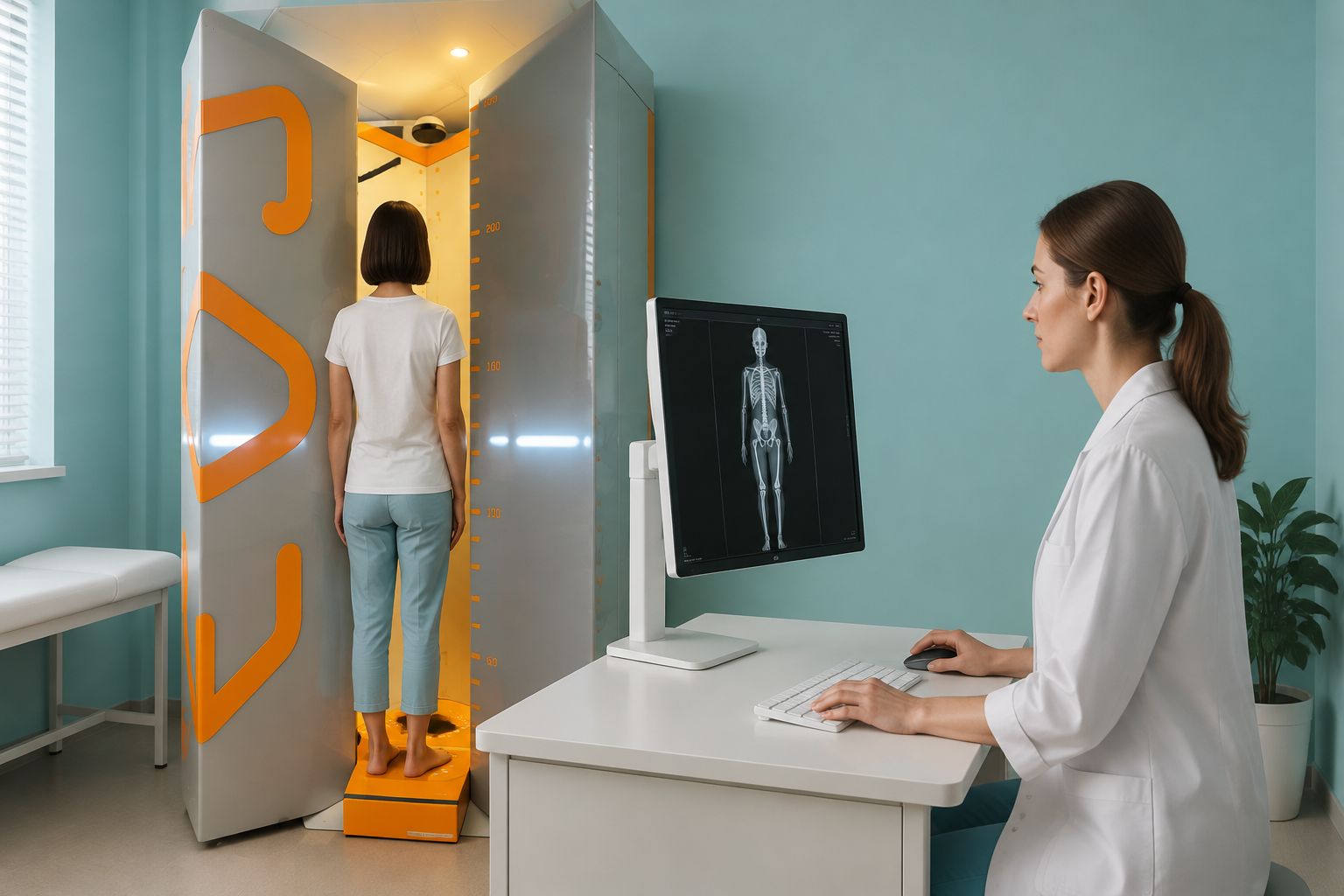
EOS Low-Dose Standing X-ray: What Is It and Why Does It Matter?
An EOS standing X-ray takes one upright image of your whole skeleton, from head to feet. It uses about 90% less radiation than a standard X-ray, around ten times less. You stand and carry your own weight during the scan. So it shows how your bones and joints really line up under gravity. Scans done lying down cannot do this.
Key points
- EOS scans you standing up and takes your whole body in one low-dose image.
- It uses about 90% less radiation than a standard X-ray, around ten times less.
- Standing imaging shows alignment problems that lying-down scans can miss.
- Pain often shows up away from its true cause. EOS helps trace the full chain.
- You do not need a referral. ScanAlign is a private, self-pay clinic at 19 Harley Street, London.
What is an EOS scan and how does it work?
An EOS scan is a type of X-ray. It images your whole skeleton in one pass while you stand naturally inside an open booth. Two very fine X-ray beams move from head to toe at the same time, one from the front and one from the side. They produce a detailed full-length image in under a minute.
The technology grew out of physics research that won a Nobel Prize. Its detectors are extremely sensitive, so they need far less radiation to make a clear image. The result is about 90% less radiation than a standard X-ray, around ten times less.
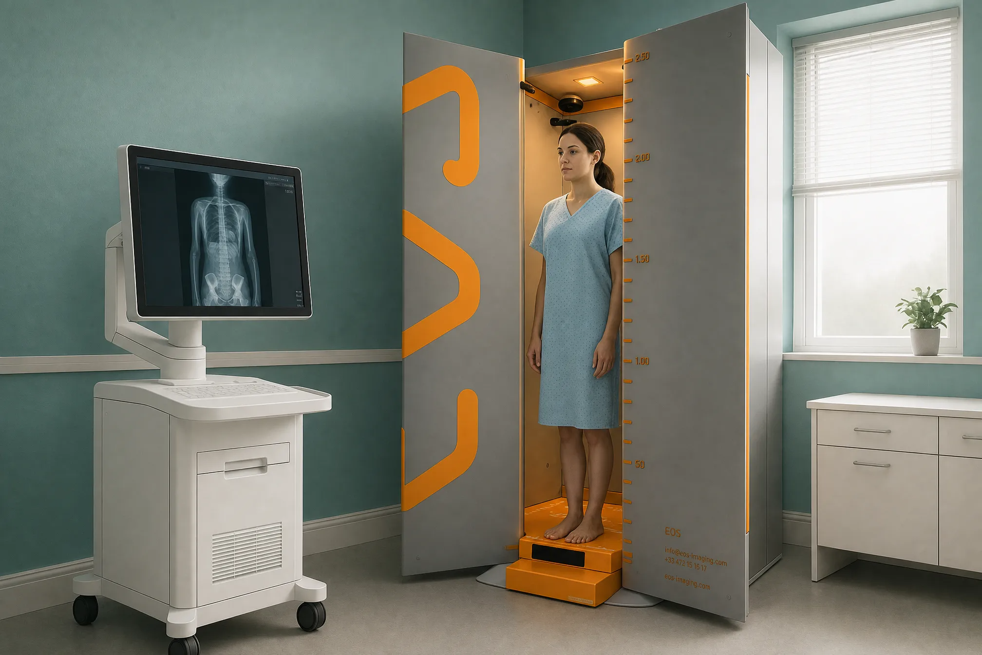
Unlike a CT scanner, the EOS booth is open and roomy. There is no tunnel or closed space, so it is comfortable even if you feel uneasy in tight spaces.
Why does standing up during an X-ray make a difference?
Standing up changes everything. When you lie down for a normal X-ray, CT or MRI, gravity no longer presses through your skeleton. Your muscles relax, your curves flatten, and your joints move into a position they are never in during daily life. The image may look reassuring, but it does not show how your body actually works.
An EOS standing X-ray images you while you carry your own weight, just as you do when you walk, stand and sit. It shows the true angles of your spine, the real tilt of your pelvis, and how your knees and hips line up under load. Some problems disappear when you lie flat, such as a small difference in leg length or a pelvis that drops to one side. On EOS, these become visible.
Would you like to know how standing imaging could help with your own problem?
Book a free video consultationWhat does an EOS scan show that other imaging misses?
An EOS scan shows the full mechanical chain of your skeleton in one image. This means your specialist can see how a problem in one area affects everywhere else. Your body is a connected system from your feet to your head. A shift at the ankle can tilt the knee, turn the hip, curve the spine and tip the shoulders. Pain often appears far from where the problem started.
See every curve in your spine while gravity and your muscles hold it in its natural position.
Find a pelvic tilt or rotation that only shows when you are upright and carrying your weight.
Measure how your knees line up over your feet and spot differences in leg length under load.
Trace how a shift in one joint forces other joints to compensate, and find the real source.
Imaging of one area, such as an X-ray of just your knee, shows only that one joint on its own. It cannot show that the knee pain might be driven by a turned hip above or a flat foot below. EOS gives the complete picture.
When is MRI the better choice? EOS is excellent for bones, joints and alignment. For soft-tissue problems, such as disc injuries, torn ligaments or nerve compression, MRI is usually the better tool. Your specialist can advise which scan you need, or whether you might benefit from both.
Is an EOS X-ray safe and how much radiation is involved?
Yes. An EOS scan uses about 90% less radiation than a standard X-ray, around ten times less. That makes it one of the lowest-dose skeletal imaging options available. The scan is quick and painless, and nothing enters your body.
The low dose also makes it practical for follow-up scans if your clinician needs to check changes over time, without building up unnecessary exposure.
What happens during an EOS scan at ScanAlign?
The process is simple. Here is what a typical visit looks like.
Free video consultation
You speak with a specialist radiographer from home. They listen to your symptoms, go over your history and confirm whether an EOS scan is the right next step.
Arrive at 19 Harley Street
You come to the clinic in central London. No preparation is needed. You can eat and drink as normal beforehand.
Stand in the scanner
You step into the open EOS booth and stand still for a short time while the two beams scan from head to toe. No tunnel, no noise, no injections.
Receive your report
A specialist reviews your images. You receive a detailed report that you can share with your consultant, surgeon or physiotherapist back home.
You do not need a referral from your doctor. ScanAlign is a private, self-pay clinic. For patients travelling from abroad, the price is confirmed during the free video consultation, and you can read more on the FAQs page.
Who should consider an EOS standing X-ray?
An EOS scan is most useful for adults with long-standing or unexplained back, hip, knee or posture problems that other scans have not fully explained. It is a diagnostic imaging tool, not a treatment. But it often provides the missing piece that guides better treatment decisions elsewhere.
- Persistent back, hip or knee pain without a clear diagnosis
- A posture concern or a body that looks uneven
- Earlier scans that showed "nothing wrong", but you still have symptoms
- A planned joint replacement or spine procedure that needs alignment data
- A need for low-dose follow-up scans over time
- Soft-tissue injuries such as disc tears, ligament damage or nerve problems (MRI is better)
- Recent fractures or injuries (go to an emergency department)
- Internal organ conditions not related to the skeleton
If you have sudden numbness, weakness, or loss of bladder or bowel control, please get urgent medical help from your doctor or the nearest emergency department rather than booking a scan.
What areas of the body can EOS imaging assess?
EOS takes your full skeleton in one standing image, so it is useful for a range of bone, joint and muscle concerns. Below are the areas where alignment imaging adds the most value.
Before you start a treatment, or change the one you are on, it is worth getting a detailed diagnosis. Knowing whether the cause sits in the pelvis, the legs or the spine changes what will actually help.
Scoliosis, lordosis, kyphosis and posture problems assessed under natural weight-bearing.
Learn more → Hip Hip alignment and pelvic tiltHip position, pelvic balance and alignment checks before or after surgery.
Learn more → Knee Knee and leg alignmentMechanical axis measurement, knock knees, bow legs and planning before knee replacement.
Learn more →Frequently asked questions about EOS standing X-rays
An EOS scan takes a full-length image of your skeleton while you carry your own weight. It is especially useful for assessing spinal alignment, pelvic balance, and the position of the legs or knees. It helps clinicians understand how your whole body works together under gravity.
An EOS scan uses about 90% less radiation than a standard X-ray, around ten times less. This makes it one of the lowest-dose skeletal imaging methods available.
No. ScanAlign is a private, self-pay clinic. You can contact us directly without a letter from your doctor.
A normal X-ray usually images one body part at a time, often while you are lying down. An EOS scan takes your entire skeleton in one standing image with far less radiation. So it shows how your bones and joints line up under your own body weight.
They answer different questions. EOS is excellent for bones, joints and whole-body alignment. MRI is usually the better tool for soft-tissue problems such as disc injuries, ligament tears or nerve compression. In some cases, both scans together give the most complete picture.
The scan itself takes under a minute. Your whole visit, including positioning and any questions, is usually quite short. No special preparation is needed.
For patients travelling from abroad, the price is confirmed during the free video consultation. You can also visit the FAQs page.
When you lie down, gravity no longer presses through your skeleton. Your muscles relax, curves flatten and joint positions change. A standing X-ray shows how your body actually carries load. It reveals alignment problems that lying-down scans can miss entirely.
No. The EOS booth is open and roomy, nothing like an MRI tunnel. You simply stand inside while the scan runs. Most people find it very comfortable.
ScanAlign is at 19 Harley Street, London, within The Harley Street Hospital. It is easy to reach by public transport, with several London Underground stations nearby.
Ready to see the full picture?
Speak with a specialist radiographer in a free video consultation from home. No referral needed, no obligation.
Book a free video consultationSources
- Deschênes S, et al. "Diagnostic imaging of spinal deformities: reducing patients' radiation dose with a new slot-scanning X-ray imager." Spine, 2010. Published by Lippincott Williams & Wilkins.
- Melhem E, et al. "EOS biplanar X-ray imaging: concept, developments, benefits, and limitations." Journal of Children's Orthopaedics, 2016;10(1):1-14.
- NHS. "X-ray: How it is performed." NHS.uk. Available at: nhs.uk/conditions/x-ray.
Written by Abbas Dhami (Specialist Diagnostic Radiographer)
This page is general information and not a diagnosis.
ScanAlign is the trading name of CoreMed Solutions Ltd and operates under The Harley Street Hospital's CQC licence.