
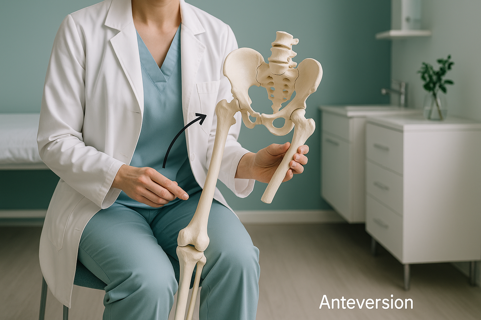
Anteversion vs Retroversion: What Is the Difference and Why Does It Matter?
Anteversion means a bone angles forward more than normal. Retroversion means it angles backward. Both most often affect the femur (thigh bone) or the hip socket (acetabulum). When the rotation is outside the typical range, it changes how you stand, walk and load your joints. This often causes pain well away from the hip itself.
Key points
- Anteversion tilts a bone forward. Retroversion tilts it backward.
- Both change how force travels through the hip, knee and lower back.
- Lying-down scans relax the muscles and can hide the real rotation.
- A standing, whole-body EOS image shows bone rotation while you carry your own weight naturally.
- Accurate measurement helps clinicians plan the right approach, whether that is physiotherapy, orthotics or surgery.
What is anteversion and how is it different from retroversion?
Anteversion describes the natural forward twist of a bone compared with another part of the skeleton. In the hip, femoral anteversion means how much the top of the thigh bone (the femoral neck) angles forward compared with the knee. Acetabular anteversion means the same forward angle of the hip socket.
Retroversion is the opposite. The bone or socket faces further backward than expected. Neither word describes a disease. They describe a direction and amount of rotation. This may or may not cause problems, depending on the person.
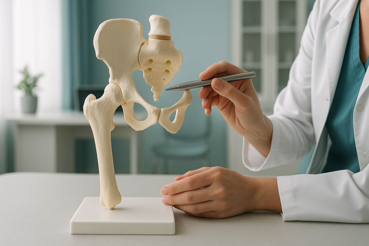
Everyone has some femoral anteversion. It is part of how the hip develops. In adults, the typical range is roughly 10 to 25 forward. When a person falls well outside that range, the way force travels through the leg changes. This can affect walking pattern (gait), how the knee tracks and how the lower back moves.
What are the signs of abnormal anteversion or retroversion?
Many people with unusual bone rotation have no symptoms at all. Others notice problems that seem to have nothing to do with the hip. Here are common patterns.
Too much anteversion often makes the feet turn inward. Retroversion can do the opposite.
The joint may sit at an angle that increases wear. This causes an ache that comes on slowly during activity.
A rotation mismatch higher up changes how the kneecap tracks. This often causes pain at the front of the knee.
The pelvis may make up for rotation differences. This puts uneven load on the lower (lumbar) spine.
Your body is a connected chain from your feet to your head. So a rotation problem at the hip can cause pain in the knee, the lower back, or even the other side. This is why a scan of one area sometimes finds nothing wrong, while the real cause sits somewhere else in the chain.
What causes too much anteversion or retroversion in adults?
In most adults, the amount of rotation is set during growth and does not change. Some people simply develop at the higher or lower end of the range. Other factors can also play a part.
The most common reason. The femur or socket simply grew with more or less twist than average.
A healed thigh bone fracture may change the rotation, especially if it was not fully corrected.
Wear-and-tear changes can shift how the joint sits. Over time, this changes its working angle.
Acetabular anteversion and retroversion matter just as much as femoral version. The two together, sometimes called "combined version", decide how well the ball fits into the socket through a full range of movement. A mismatch between the two can cause impingement or instability, even when each measurement on its own looks acceptable.
How is anteversion checked with a standing scan?
A standing, whole-body EOS image captures both legs and the pelvis in one calibrated view while you carry your own weight. This matters because lying-down scans relax the muscles around the hip. The leg can then fall into a resting position that may hide the true rotation.
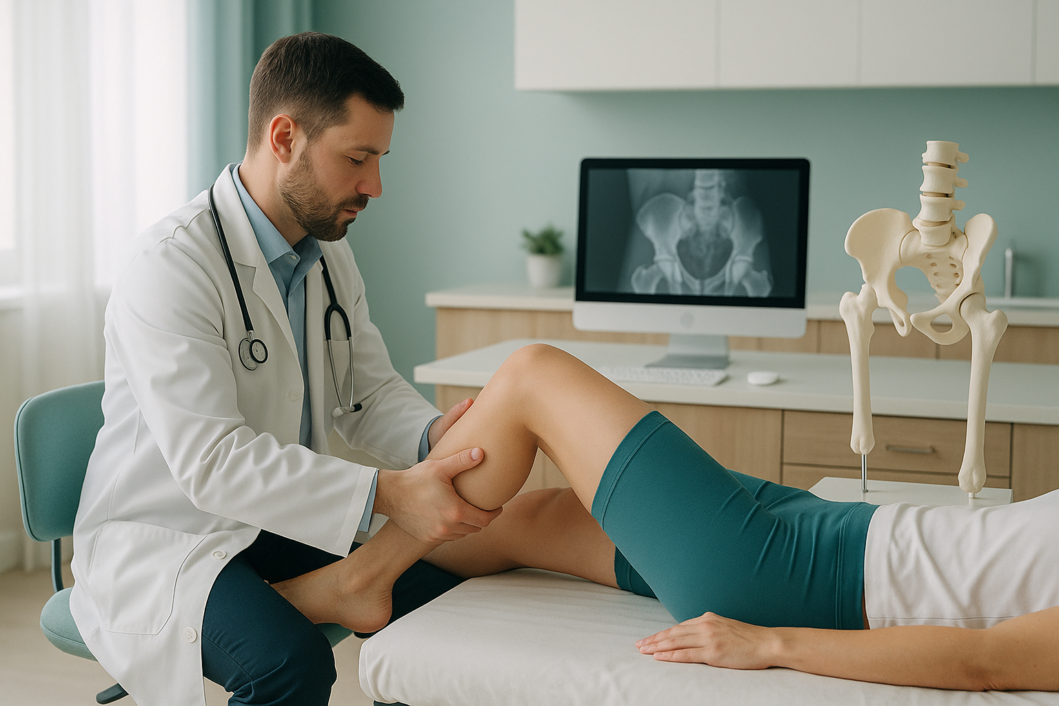
When you stand, your muscles switch on and your pelvis settles into its working position. The image captures how the femur and the socket line up as they really work in daily life. A full-length view also shows how the knees and feet respond to that rotation. It reveals the whole chain of compensation in one image.
EOS uses about 90% less radiation than a standard X-ray, around ten times less. That matters because rotation checks sometimes need several views, and a lower dose means less total exposure.
Want to understand how your hip rotation affects the rest of your alignment? Speak with our team first, from home, at no cost.
Book a free video consultationWhat can be done about abnormal anteversion or retroversion?
The right approach depends entirely on what the scan shows, how the rotation works with the rest of your alignment, and whether it is causing symptoms. ScanAlign provides the diagnostic imaging, not the treatment. Your treating specialist, whether an orthopaedic surgeon, physiotherapist or sports medicine specialist, uses the results to decide the next steps.
Common options include targeted physiotherapy to strengthen the muscles around the hip, retraining how you walk, orthotic support, or, in more serious cases, surgical correction such as a derotational osteotomy. All of these benefit from precise rotation measurements taken while you carry your own weight.
So before you start treatment, or change the one you are on, it is worth getting a detailed diagnosis. Knowing whether the cause sits in the pelvis, the legs or the spine changes what will actually help.
When MRI is the better choice: EOS shows bones, joints and alignment clearly. For soft-tissue concerns such as labral tears, cartilage damage or nerve involvement, MRI is usually the better tool. If your treating specialist suspects a soft-tissue cause, they may recommend MRI alongside or instead of a standing bone assessment.
Frequently asked questions about anteversion and retroversion
Femoral anteversion is the forward twist of the top of your thigh bone compared with the knee. Everyone has some of it. When it is greater than the typical range, the whole leg tends to turn inward. This can affect how you walk and how your knee tracks.
Anteversion means a bone or socket faces further forward than average. Retroversion means it faces further backward. They are opposite ends of the same range of rotation, and both can change how the hip joint works.
Yes. When the femur is rotated more than usual, the kneecap can track differently. This changes how force moves through the knee. It can cause pain at the front of the knee, even when the knee itself has no structural damage.
The bone rotation itself usually stays stable once growth is complete. But the symptoms it causes can get worse as joint wear increases or as muscle strength declines with age. That is why accurate measurement becomes more important over time.
When you lie down, your muscles relax and gravity pulls the leg into a resting position. This can hide the true rotational alignment. Standing switches on the muscles and loads the joints. So the image captures how your skeleton actually works during daily activities.
Acetabular anteversion describes how far forward the hip socket faces. Together with femoral version, it decides how well the ball sits in the socket. A mismatch between the two can cause impingement or instability.
No. You can book directly yourself. The process starts with a free video consultation. Our team discusses your situation, confirms that a standing scan is suitable, and explains what the session involves.
EOS uses about 90% less radiation than a standard X-ray, around ten times less. This is especially helpful when several views or follow-up scans are needed.
The price for patients travelling from abroad is confirmed during the free video consultation. You can also read our FAQs page.
The bone rotation itself cannot be changed without surgery. However, physiotherapy, strengthening and changes to how you walk can often manage symptoms well. Accurate imaging helps your treating specialist decide whether non-surgical care is likely to be enough or whether a surgical opinion is worthwhile.
See how your alignment really works
A standing, whole-body image can reveal the rotation that scans of one area miss. Start with a free video consultation before you plan your trip to London.
Book a free video consultation Learn more about EOS imagingSources
- Tönnis D, Heinecke A. "Acetabular and femoral anteversion: relationship with osteoarthritis of the hip." The Journal of Bone and Joint Surgery (American), 1999;81(12):1747-70.
- Dubousset J et al. "EOS: a new imaging system with low dose radiation in standing position for spine and bone & joint disorders." Journal of Musculoskeletal Research, 2010;13(1):1-12.
- NHS (UK National Health Service). "Hip pain in adults." NHS website, reviewed 2023. nhs.uk/conditions/hip-pain
Written by Abbas Dhami (Specialist Diagnostic Radiographer)
This page is general information and not a diagnosis.
ScanAlign is the trading name of CoreMed Solutions Ltd and operates under The Harley Street Hospital's CQC licence.