
Spinal Fusion: What Happens During Surgery, and What Is Recovery Really Like?
Spinal fusion joins two or more vertebrae for good, so they heal into one solid bone. The surgeon uses screws, rods and sometimes a bone graft or cage to hold the vertebrae in place while they fuse. Recovery usually takes several months, and you return to daily activities step by step. You will lose some range of movement at the fused levels.
Key points
- Spinal fusion locks vertebrae together to stop painful movement at a damaged segment.
- The operation carries real risks, including infection, nerve injury and adjacent segment disease.
- It can take three to twelve months before you feel the full benefit.
- Standing, weight-bearing imaging before and after surgery helps your surgeon assess the alignment of your whole spine, not just the fused area.
- EOS imaging uses about 90% less radiation than a standard X-ray, around ten times less.
What is spinal fusion and why is it done?
Spinal fusion is an operation that joins two or more vertebrae in your spine for good. The aim is to stop movement at a painful or unstable segment. Your surgeon may suggest it when treatments without surgery, such as physiotherapy, injections or medicines, have not given enough relief.
Common reasons for lumbar fusion surgery include severe disc degeneration, a vertebra that has slipped forward or backward (spondylolisthesis or retrolisthesis), fractures of the spine, or instability after a laminectomy.
In time, the fused vertebrae grow together into one piece of bone. This stops movement at that level, and so stops the friction and pain there. But it also takes away some flexibility from your spine. That is why the decision to go ahead is always weighed carefully.
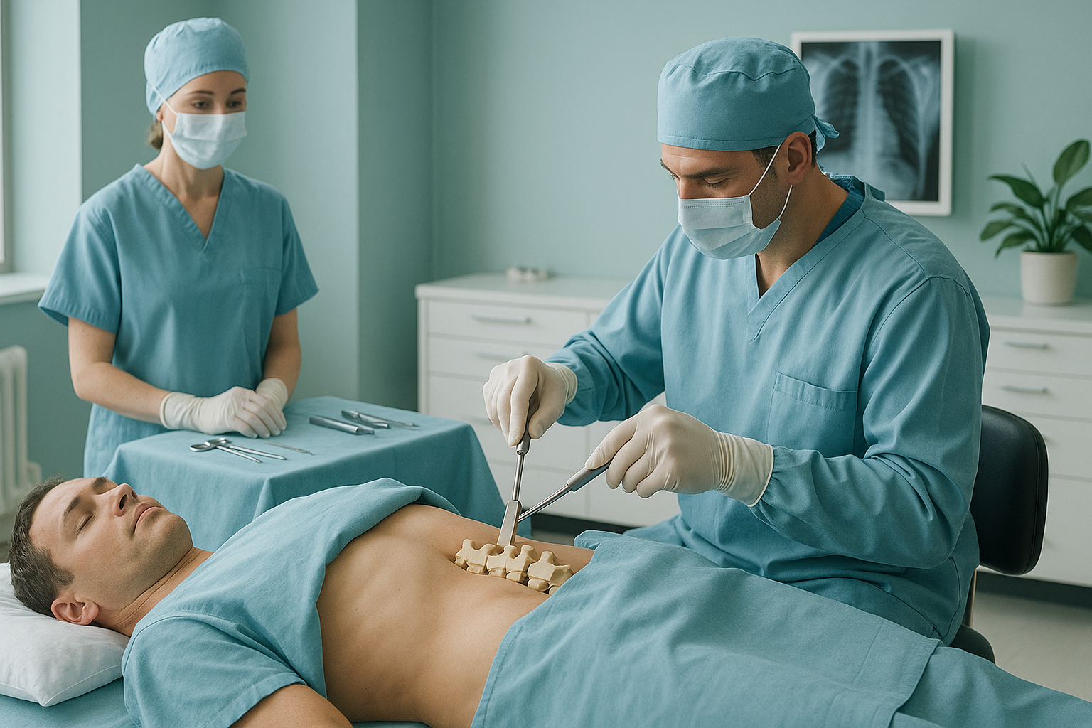
What actually happens during spinal fusion surgery?
The surgeon reaches the spine from the back (posterior), the front (anterior) or sometimes both. This depends on which levels need fusing and the exact problem being treated. The damaged disc material is usually removed. Then an interbody cage, a small spacer filled with bone graft material, is placed in the disc space. Pedicle screws are put into the vertebrae above and below, and rods connect the screws to hold everything rigid.
The bone graft can come from your own body, from a donor, or from a synthetic substitute. Over several months, new bone grows through and around the graft until the vertebrae fuse into one piece.
A single-level fusion usually takes two to four hours under general anaesthesia. Multi-level fusions take longer and carry higher risk. This is why surgeons try to fuse only the levels that truly need it.
What are the risks of spinal fusion?
All surgery carries risk. Spinal fusion has specific risks you should understand before you give consent. These include infection, blood clots, nerve damage, and the chance that the bones do not fuse properly (non-union). Your surgeon will discuss your own level of risk with you.
Because the fused levels no longer move, the discs and joints above and below have to work harder. Over time this extra stress can cause new problems at the neighbouring segments.
Every fused level takes away some flexibility. With a single-level fusion this is often barely noticeable. Multi-level fusions can reduce bending and twisting more.
If the fusion does not keep the spine's natural curves, you can end up leaning forward when you stand. This is called flatback syndrome and may need further surgery.
Screws can loosen, rods can shift, and cages can move out of place. This sometimes needs revision surgery. Good bone quality and correct alignment lower this risk.
Important. If you develop sudden leg weakness, numbness spreading to both legs, or loss of bladder or bowel control after spinal surgery, get urgent medical help. Contact your surgical team or go to the nearest emergency department.
What is recovery after spinal fusion really like?
Recovery after spinal fusion is gradual. Most people stay one to four days in hospital. They then recover at home for several weeks before going back to light activities. Full recovery, meaning the bone has fused solidly, usually takes anywhere from three to twelve months.
The usual recovery timeline
You will be resting at home and managing pain with prescribed medicines. Walking short distances is encouraged. Bending, lifting and twisting are restricted.
You walk a little more each day. You may start guided physiotherapy. Most people can manage light daily tasks but are not yet ready to drive or work.
Many people go back to desk-based work. Physiotherapy focuses on strengthening the core. Your surgeon will check scans to see how the fusion is progressing.
The bone keeps fusing and getting stronger. More active work and exercise can slowly start again. Some stiffness or discomfort may remain but should improve steadily.
Life after spinal fusion is different for everyone. Some people feel much better once the source of pain is stabilised. Others find the loss of flexibility takes getting used to. Realistic expectations, guided rehabilitation and careful planning before surgery all improve outcomes.
Thinking about surgery or already had it? A standing spine scan can show your surgeon how your whole spine is aligned under your own body weight. This matters for planning and follow-up.
Book a free video consultationWhy does standing imaging matter before and after spinal fusion?
Standing imaging matters because your spine behaves differently when you are upright and bearing weight than when you are lying down. A conventional X-ray, CT or MRI is taken while you lie flat. In that position, your muscles relax, your curves change, and gravity is no longer loading your joints. The image can look reassuringly normal, even when you have significant alignment problems when standing.
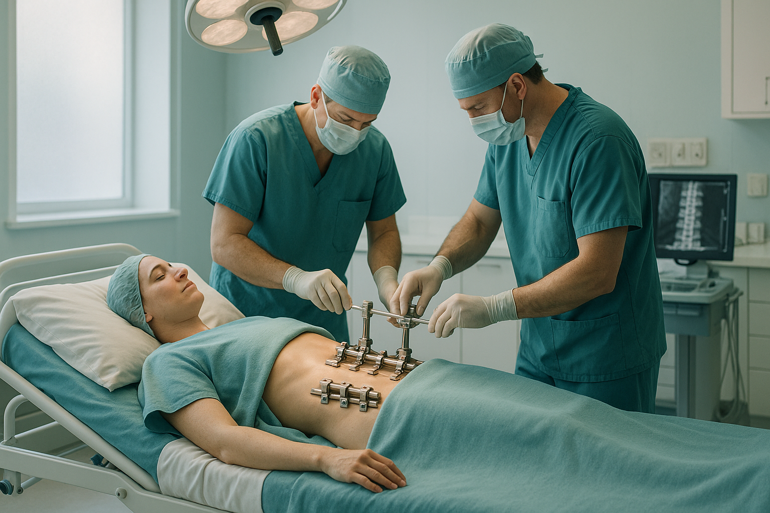
This is especially important for spinal fusion. Your surgeon needs to understand your sagittal balance, the front-to-back alignment of your whole spine when upright. If the fusion is planned at the wrong angle, or without taking your overall balance into account, you could end up leaning forward, developing flatback syndrome, or putting too much stress on the segments above and below the fusion.
ScanAlign standing imaging uses EOS technology to capture your full spine and legs in a single standing image, with about 90% less radiation than a standard X-ray, around ten times less. This gives your surgical team a complete picture of how your body is aligned in real-life conditions.
When MRI is the better choice. EOS is excellent at showing bones, joints and whole-body alignment. For soft tissue detail, such as disc bulges, nerve compression or spinal cord problems, MRI is usually the better tool. Many people benefit from both.
Who benefits from standing imaging around spinal fusion?
A standing whole-body scan is most useful at two key moments. The first is before surgery, when your surgeon is planning the fusion levels and angles. The second is after surgery, when checking that the fusion is holding proper alignment under gravity.
- Are being considered for spinal fusion and want a clear starting picture of your alignment
- Have had fusion surgery and still feel off-balance or in pain
- Need follow-up imaging with lower radiation exposure
- Want your surgeon to see your full sagittal profile standing up
- You need soft-tissue detail (MRI is usually better for this)
- You need an emergency scan for an injury or sudden severe symptoms
- You cannot stand still for the short scanning time
Before you start a treatment, or change the one you are on, it is worth getting a detailed diagnosis. Knowing whether the cause sits in the pelvis, the legs or the spine changes what will actually help.
Frequently asked questions about spinal fusion
A single-level fusion usually takes two to four hours. Multi-level or more complex fusions may take longer. Your surgeon will give you a more accurate estimate based on your own case.
There is pain after the operation, which is managed with medicines in hospital and at home. This surgical pain usually improves over the first few weeks. The original back or leg pain that led to surgery often starts to ease once the fusion stabilises.
This depends on your job. People with desk-based jobs may go back after around six to twelve weeks. Physically demanding jobs may need three to six months or more. Your surgeon and physiotherapist will guide you.
When vertebrae are fused, the segments above and below take on extra movement and stress. Over time this can cause new disc degeneration or instability at those neighbouring levels. It is one of the most discussed long-term risks of fusion.
You will lose some range of movement at the fused levels. With a single-level fusion, most people barely notice the difference. Multi-level fusions reduce flexibility more noticeably. The rest of your spine and your hips make up for much of the lost movement.
Standing images show your spine under gravity, which reveals your true alignment. Your surgeon needs to see this to plan the correct angle and position for the fusion. Without it, there is a greater risk of problems like flatback syndrome or poor sagittal balance after surgery.
EOS uses about 90% less radiation than a standard X-ray, around ten times less. This is especially helpful if you need several follow-up scans after surgery to check how the fusion is progressing.
No. You can refer yourself. The process starts with a free video consultation, where a specialist radiographer reviews your situation and confirms whether a standing scan is right for you.
The price for patients travelling from abroad is confirmed during the free video consultation, which you can start from the book a standing spine scan page.
They serve different purposes. EOS shows your overall bony alignment standing up. MRI shows soft tissue detail like discs and nerves. Most fusion patients benefit from both types of imaging, as each answers a different clinical question.
Understand your alignment before or after fusion
A standing whole-body scan shows your surgeon exactly how your spine is aligned under gravity. No referral needed.
Book a free video consultationSources
- NHS. "Lumbar decompression surgery." nhs.uk. Accessed 2024.
- Glassman SD, et al. "The impact of positive sagittal balance in adult spinal deformity." Spine, 2005;30(18):2024-2029.
- Illés T, Somoskeöy S. "The EOS imaging system and its uses in daily orthopaedic practice." International Orthopaedics, 2012;36(7):1405-1411.
Written by Abbas Dhami (Specialist Diagnostic Radiographer)
This page is general information and not a diagnosis.
ScanAlign is the trading name of CoreMed Solutions Ltd and operates under The Harley Street Hospital's CQC licence.