
What Is the HKA Angle, and How Is Leg Alignment Measured?
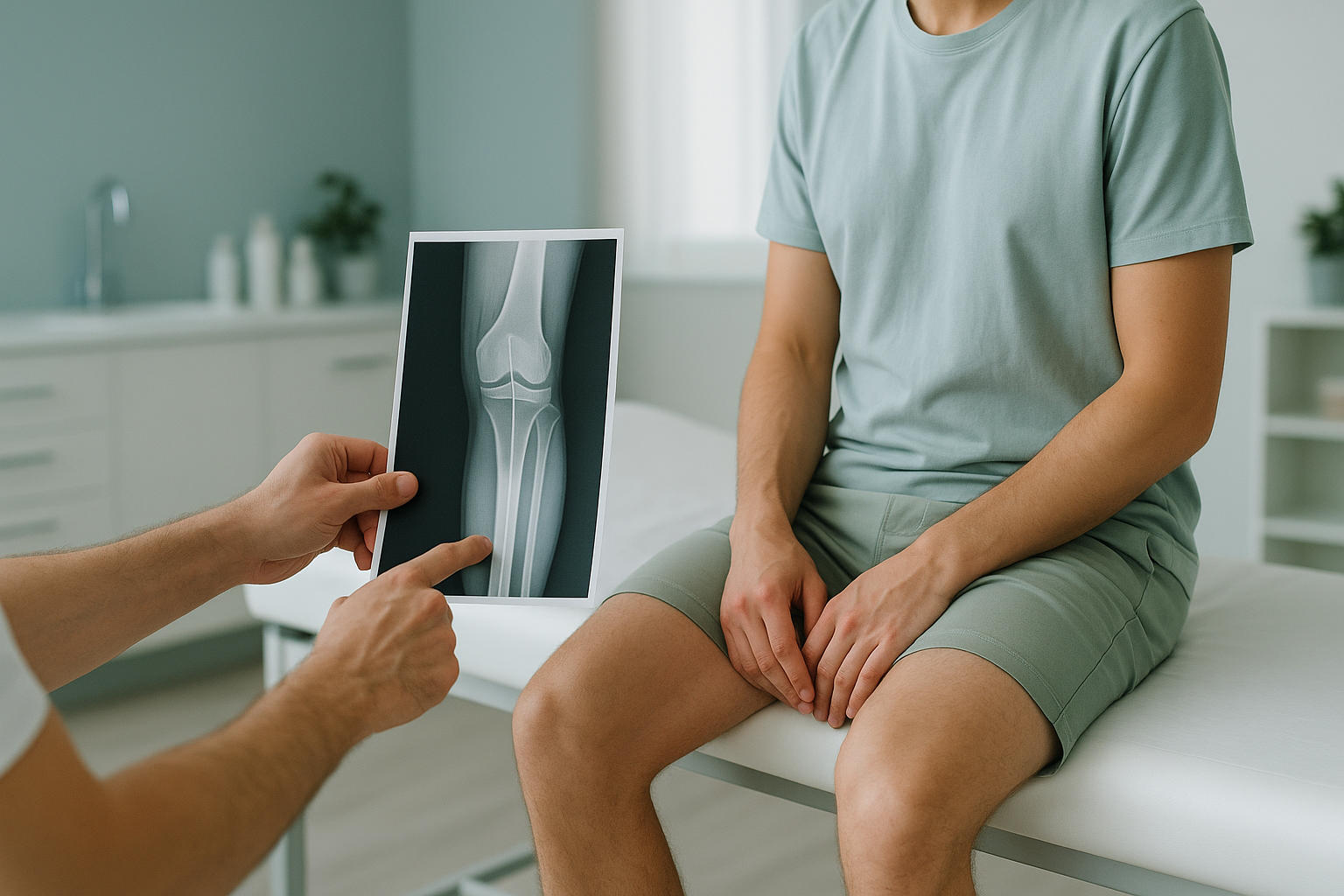
The HKA angle (hip-knee-ankle angle) is the measurement between two lines. The first runs from the centre of your hip to the centre of your knee. The second runs from the centre of your knee to the centre of your ankle. It tells clinicians whether your leg is straight, bowed inward or angled outward. To measure it accurately, you need a full-leg standing x-ray.
Key takeaways
- The HKA angle measures overall leg alignment from hip to ankle in one number.
- A straight leg has an HKA close to 180 degrees. Values above or below this point to valgus (knock-knee) or varus (bow-leg) alignment.
- Standing, weight-bearing imaging is essential, because lying-down scans can hide real alignment problems.
- The HKA angle is used to plan knee surgery, osteotomy and joint replacement, and to monitor how arthritis progresses.
- EOS imaging captures the full leg in one low-dose exposure with about 90% less radiation than a standard x-ray.
What exactly is the HKA angle?
The HKA angle is the angle formed at your knee when a clinician draws two lines on a full-length image of your leg. One line runs from the centre of the femoral head (hip ball) to the middle of the knee. The other runs from the middle of the knee to the centre of the ankle joint. Together, these lines form the mechanical axis of your leg.
In a perfectly straight leg, the HKA angle is roughly 180 degrees. Most people have a small natural variation. When the angle shifts noticeably, the load going through your knee is no longer spread evenly. One compartment of the joint takes more stress than it should, and that can speed up cartilage wear.
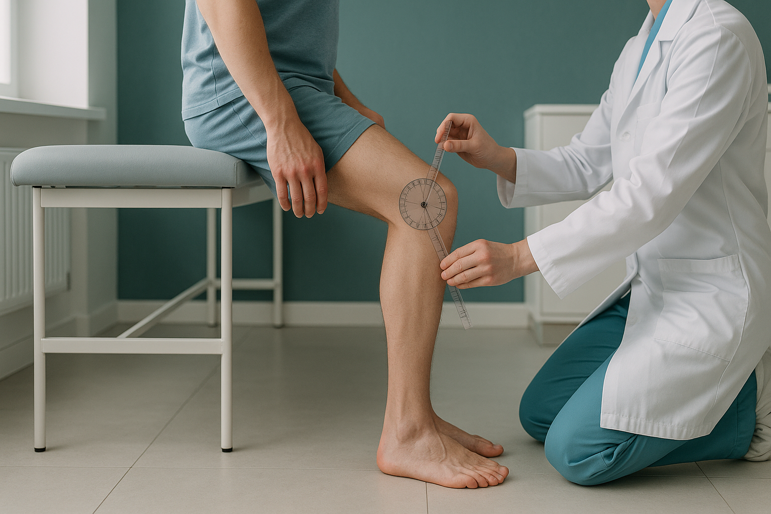
You may also hear clinicians talk about the Mikulicz line. This is a single straight line drawn from the centre of the femoral head directly to the centre of the ankle. In a well-aligned leg, the Mikulicz line passes through the middle of the knee. When alignment is off, the line shifts to one side. The distance between where it lands and the centre of the knee is called the mechanical axis deviation.
How is leg alignment measured?
Leg alignment is measured with a full-length radiograph that captures the hip, knee and ankle in one image. The radiographer marks the centre of each joint, and the software calculates the HKA angle and the mechanical axis deviation. This is sometimes called a scanogram or a long-leg view.
Without proper calibration, magnification error can distort measurements. Standing EOS imaging uses a fixed, known distance to reduce this.
A knee-only x-ray cannot show the HKA angle. The image must include the hip and ankle to map the full mechanical axis.
If the HKA angle is below 180, the knee angles inward (varus, or bow-leg). Above 180 means the knee angles outward (valgus, or knock-knee).
Older methods join several smaller x-rays together, which can create errors where the images meet. Modern systems like EOS capture the whole leg in one continuous sweep. This removes the joining problem and gives a more reliable measurement.
Why does leg alignment need to be measured standing up?
Standing imaging shows your true alignment under load. When you lie down for a CT scan or a standard x-ray, gravity no longer presses your joints together, your muscles relax, and small alignment shifts disappear. The image looks straighter than reality.
Your full body weight presses through your joints. This reveals any bowing, tilting or rotation. The HKA angle from a standing image reflects what your knee actually goes through when you walk and in daily life. This is the measurement surgeons use for osteotomy planning and surgical templating.
Without your body weight pressing down, ligaments and soft tissue relax. A mildly bowed leg can look nearly straight, and the HKA angle reading may underestimate the real problem. Lying-down scans are still useful for other things, but they are not reliable for measuring weight-bearing alignment.
Your body is a connected chain from your feet to your head. If alignment is off at the knee, the hip and ankle compensate. A standing image captures this whole chain, so the clinician can see where the problem starts and where it is felt. Pain in your knee, for example, may partly come from a pelvis that is not level, something a knee-only scan would never show.
Why does the HKA angle matter for your knee?
The HKA angle matters because it decides where load falls across your knee joint. An even load means even wear. A shifted load means the cartilage on one side breaks down faster. This is a common driver of osteoarthritis in the inner or outer compartment of the knee.
Tracking the HKA angle over time shows whether alignment is getting worse and how fast cartilage loss is progressing.
Surgeons use the HKA angle for osteotomy planning, to decide exactly how much bone to correct, and for templating knee replacements.
After surgery, a follow-up HKA measurement confirms that the corrected alignment is holding as expected.
Beyond surgery, the HKA angle helps physiotherapists and sports medicine specialists understand why you have persistent knee pain, why one side wears out faster, or why you keep injuring the same area. It gives a measurable, objective baseline rather than a guess.
What are the signs you might need your HKA angle measured?
You may benefit from an HKA measurement if you have persistent or unexplained knee pain, visible bowing or knock knees, or if a surgeon has recommended alignment imaging before a procedure.
- Ongoing pain on the inner or outer side of the knee with no clear cause on standard imaging
- Visible bow legs or knock knees that seem to be getting worse
- A recommendation for osteotomy or knee replacement
- Past knee surgery and continued problems
- Pain in the hip or ankle that may be linked to leg alignment
- Sudden severe knee pain after an injury, with swelling and being unable to put weight on the leg
- Numbness, weakness or tingling spreading down the leg
- Loss of bladder or bowel control (go to the nearest emergency department immediately)
When MRI is the better choice: EOS and standing x-rays show bones, joints and alignment very well. For soft tissue problems such as ligament tears, meniscus damage or nerve compression, MRI is usually the better tool. Your clinician can advise which scan is right for you.
How is the HKA angle assessed with a standing EOS scan?
An EOS scan captures your full skeleton from head to ankle while you stand naturally. It uses about 90% less radiation than a standard x-ray, around ten times less. The image is taken in one continuous pass, so there are no joining errors. The calibration is built into the system, which reduces magnification error.
To learn more about the technology, read about what an EOS scan is. The process is similar to Cobb angle measurement for the spine. Both rely on a full-length, weight-bearing image to get an accurate reading.
At ScanAlign standing imaging, your scan is reviewed by a specialist radiologist. They measure the HKA angle, mechanical axis deviation and related alignment values, then give you a detailed report your clinician can use.
Book a free video consultationYou do not need a referral from your doctor. You can book a measured alignment scan directly and take the results back to your surgeon, physiotherapist or orthopaedic consultant at home.
Before you start treatment, or change the treatment you are on, it is worth getting a detailed diagnosis. Knowing whether the cause sits in the pelvis, the legs or the spine changes what will actually help.
Frequently asked questions about the HKA angle
HKA stands for hip-knee-ankle. It describes the angle formed where the mechanical axis of the thigh bone (femur) meets the mechanical axis of the shin bone (tibia) at the knee.
A perfectly straight leg has an HKA angle of about 180 degrees. Most people fall within a small range either side of that. Your clinician will read your result in the light of your symptoms and activity level.
Varus alignment means the knee angles inward, giving a bow-legged look. Valgus alignment means the knee angles outward, resulting in knock knees. The HKA angle tells you which pattern is present and by how much.
No. A standard knee x-ray only captures the knee joint itself. You need a full-length image that includes the hip and ankle, so the mechanical axis can be drawn and the angle calculated.
When you lie down, gravity is no longer pressing your joints together and your muscles relax. This can hide real alignment shifts. Standing imaging shows the alignment your joints go through during everyday activities like walking and standing.
Yes. EOS uses about 90% less radiation than a standard x-ray, around ten times less. It captures the full leg in a single low-dose sweep, which is especially helpful when repeat measurements are needed over time.
No. ScanAlign accepts self-referrals. You can book directly and share the results with any clinician involved in your care.
The Mikulicz line is a straight line drawn from the centre of the hip to the centre of the ankle. In a well-aligned leg it passes through the middle of the knee. If it falls to one side, the distance from the knee centre is called the mechanical axis deviation.
The price for patients travelling from abroad is confirmed during the free video consultation. In that call, you can also discuss whether an alignment scan is right for you.
Surgeons use the HKA angle for pre-operative templating. It helps them decide the size and position of the implant, and whether any extra correction is needed to restore balanced alignment across the joint.
Wondering about your leg alignment?
A free video consultation lets you talk through your symptoms with a specialist radiographer from wherever you are, and find out whether a standing alignment scan in London is right for you.
Book a free video consultationWritten by Abbas Dhami (Specialist Diagnostic Radiographer)
Sources
- Paley, D. Principles of Deformity Correction. Springer, 2002. (Foundational text on mechanical axis and alignment measurement.)
- Cooke, T.D.V. et al. "Radiographic assessment of bony contributions to knee deformity." Clinical Orthopaedics and Related Research, 1997; 344: 28–33.
- EOS imaging clinical documentation. "Low-dose biplanar radiography for orthopaedic applications." Available from EOS imaging (now part of Alphatec Holdings).
This page is general information and not a diagnosis.
ScanAlign is the trading name of CoreMed Solutions Ltd and operates under The Harley Street Hospital's CQC licence.