
What Imaging Does a Surgeon Need to Plan a Hip Replacement?
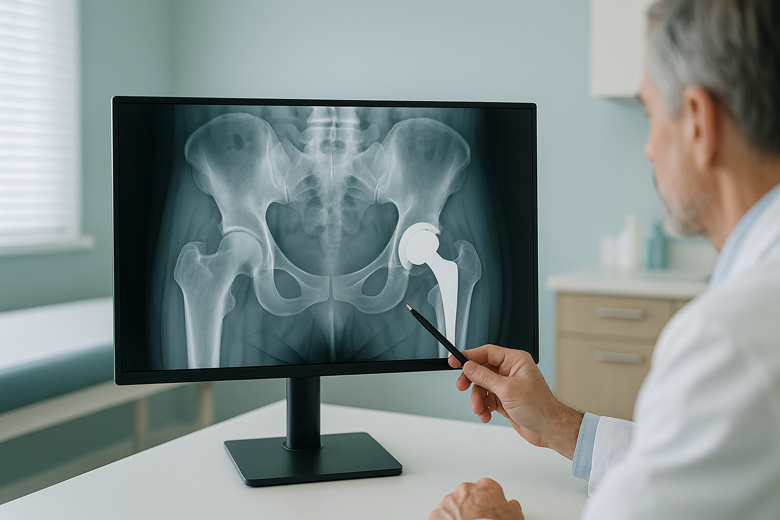
To plan a hip replacement accurately, your surgeon needs a calibrated, weight-bearing x-ray of the whole pelvis and both hips. Standing imaging shows your true pelvic tilt, any leg length difference and how your joints line up under load. All of these affect implant size, cup position and the overall surgical plan.
Key points
- Surgeons rely on calibrated x-rays to template the correct implant size and position.
- Lying-down images can hide pelvic tilt. This makes cup angle and leg length measurements less reliable.
- A standing, whole-body image shows the pelvis and spine together. It gives a full picture of your alignment.
- An EOS standing x-ray gives a non-magnified, life-size image with about 90% less radiation than a standard x-ray.
- You do not need a referral from your doctor for a private scan before surgery.
What x-rays does a surgeon need before a hip replacement?
At the least, a surgeon needs an anteroposterior (front-on) x-ray of the full pelvis and a lateral (side-on) view of the affected hip. These images are used for surgical templating. This means placing implant outlines over your x-ray on a computer, to choose the best size and position for the new parts.
For templating to work, the image must be calibrated. This means the x-ray includes a metal ball of a known size placed beside your hip, so the software can correct for any magnification. If magnification is not taken into account, the implant size can be wrong by a clinically significant amount.
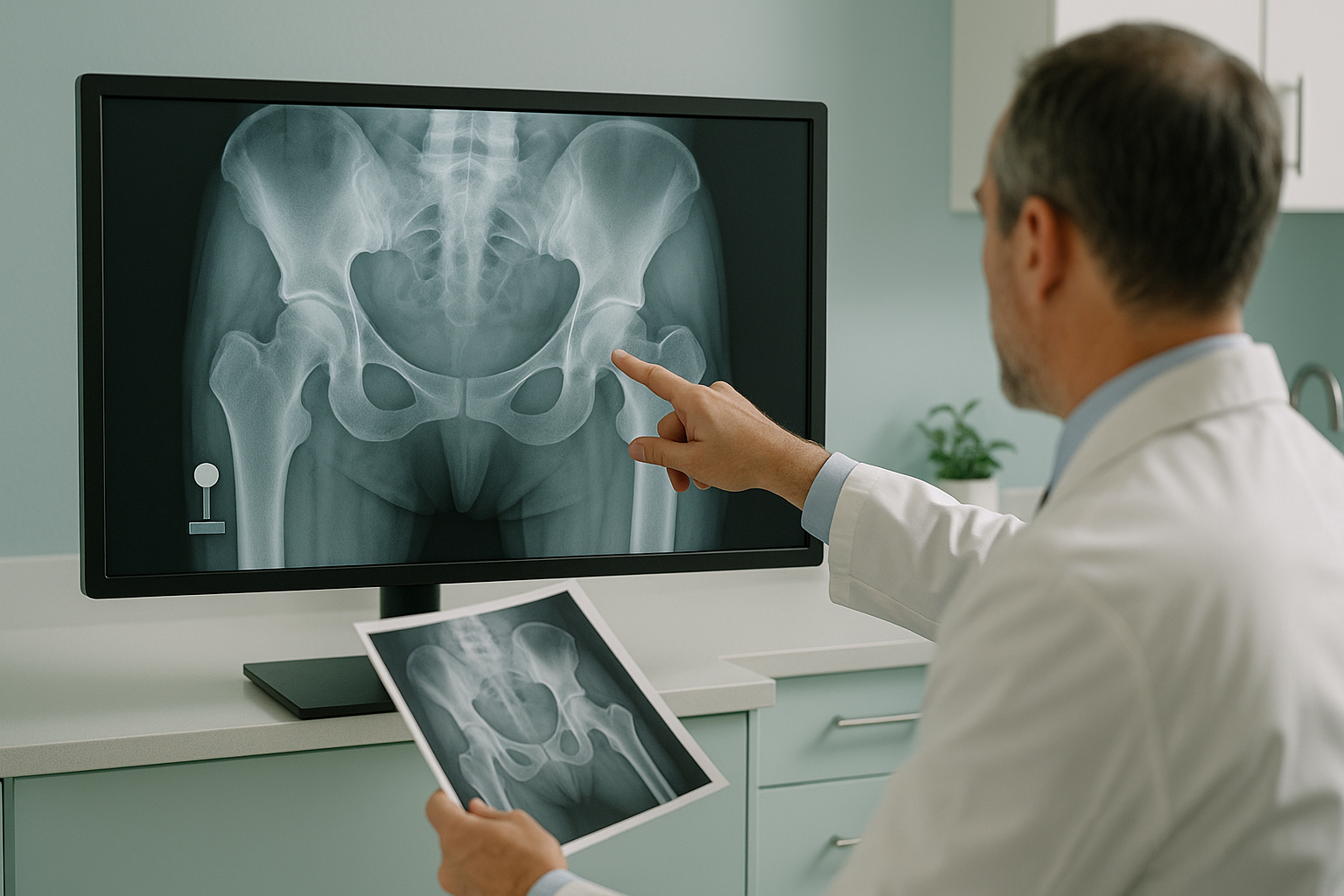
The main measurements your surgeon takes from these images are:
- Implant size. The femoral stem and acetabular cup must fit your anatomy precisely.
- Cup angle. The angle at which the socket part is placed affects stability and wear.
- Offset. The distance from the centre of the hip to the shaft of the femur. It controls muscle tension and the way you walk.
- Leg length. Your surgeon plans to restore equal leg lengths, so an accurate starting measurement is essential.
Why does standing imaging matter for hip replacement planning?
Standing imaging matters because your pelvis tilts differently when you are upright and bearing weight than when you lie down. On an x-ray table, your muscles relax and your pelvis falls into a neutral position. The image may not show the alignment you actually live with.
Pelvic tilt has a direct effect on the angle of the acetabular cup. A cup that looks well placed on a lying-down image can be too steep or too flat once you stand up. This is part of a wider assessment called spinopelvic analysis, which looks at how your spine and pelvis work together.
Shows your real, weight-bearing alignment. Your pelvic tilt, any leg length difference and the natural curve of your spine are all captured as they are in daily life. The image is true life size with no magnification error, so there is no need to guess a correction factor.
Muscles relax. Alignment shifts. With no gravity loading the joint, the pelvis can tilt forward or backward compared with standing. Magnification varies with your body size and your distance from the x-ray source. This means the calibration marker may not exactly match the plane of your hip joint.
If you have a stiff or fused spine, your pelvis compensates more than usual when you move between sitting and standing. A standing image makes that compensation visible. It gives your surgeon the information needed to place the cup safely. Conditions such as developmental dysplasia of the hip or unusual hip anteversion and retroversion also benefit from this fuller picture.
What is included in a hip scan before a hip replacement?
A hip imaging session before surgery at ScanAlign, private standing imaging in London, includes everything your surgeon needs for templating and alignment assessment. For full details, see our hip replacement imaging service.
A head-to-foot or pelvis-to-ankle image taken while you stand naturally.
No calibration ball needed. The image is at a 1:1 scale, which removes magnification error.
Pelvic tilt, sacral slope and spinal balance are all visible on the same image.
A report from a specialist radiographer, with images formatted for surgical templating software.
How does an EOS standing x-ray compare with a standard hip x-ray?
An EOS standing x-ray captures the full skeleton in one pass while you stand upright. Below is a quick comparison with a conventional hospital x-ray for hip replacement planning.
| Feature | Standard x-ray | EOS standing x-ray |
|---|---|---|
| Patient position | Usually lying down | Standing, weight-bearing |
| Magnification | Present. Needs calibration ball | Non-magnified, true 1:1 scale |
| Pelvic tilt visible | Not under real-world loading | Yes, under natural weight-bearing |
| Radiation dose | Standard | About 90% less, around ten times less |
| Full spine included | Separate image needed | Captured in the same scan |
Before you start treatment, or change the one you are on, it is worth getting a detailed diagnosis. Knowing whether the cause sits in the pelvis, the legs or the spine changes what will actually help.
How do I book imaging before a hip replacement?
You do not need a referral from your doctor. You can book yourself, or bring a request from your orthopaedic surgeon.
Free video consultation
A short call to talk about your surgery timeline and confirm the scan is right for you.
Visit us at 19 Harley Street, London
The scan itself takes only a few minutes. You stand still in normal clothing (no metal). No preparation is needed.
Images reviewed
A specialist radiographer reviews your images and prepares a report with files ready for templating.
Sent to your surgeon
The report and images are shared with your surgical team, ready for their planning session.
Ready to get the imaging your surgeon needs? You can book hip imaging before surgery or start with a free consultation.
Book a free video consultationIs an EOS scan safe before hip replacement surgery?
Yes. An EOS scan uses about 90% less radiation than a standard x-ray, around ten times less. It is a quick, non-invasive procedure with no injections and no contrast dye. You simply stand in the scanner for a few seconds.
When is MRI better? EOS shows bones, joints and alignment clearly. If your surgeon also needs to assess soft tissue, cartilage, tendons or nerves around the hip, MRI is usually the better tool. Many patients benefit from both.
Frequently asked questions
You need a calibrated anteroposterior x-ray of the full pelvis and a lateral view of the affected hip. A standing, non-magnified image is the most accurate option for surgical templating. It shows your true pelvic tilt and leg length under load.
No. You can book a private scan yourself. You are also welcome to bring a referral from your orthopaedic surgeon if you have one.
Pelvic tilt changes the effective angle of the acetabular cup once the implant is in place. If the surgeon plans from a lying-down image where the pelvis is in a different position, the cup angle may not be optimal when you stand up. Standing imaging shows your real tilt.
A non-magnified x-ray produces images at true life size. Standard x-rays enlarge the image slightly because of how the beam spreads. A non-magnified image removes that error, so implant templates can be placed directly over it without correction factors.
An EOS scan uses about 90% less radiation than a standard x-ray, around ten times less. This is especially helpful if several images are needed during your surgical planning.
Yes. EOS images are provided in standard DICOM format and work with common digital templating systems. Because they are already at 1:1 scale, no magnification calibration step is needed.
The scan itself takes only a few minutes. You stand still inside the EOS system while it captures a full image in a single pass. Most appointments, including paperwork, are short and simple.
Wear comfortable clothing without metal fastenings (zips, buttons, underwire). Most people can be scanned in light gym-style clothing. You will be asked to remove belts, jewellery and anything metal around the scan area.
The price for patients travelling from abroad is confirmed during your free video consultation.
Yes, and it is especially valuable. A stiff or fused spine limits how much your pelvis can tilt between sitting and standing. A standing image captures your actual pelvic position, which helps your surgeon choose a safer cup angle.
Get the imaging your surgeon needs
A standing, non-magnified image gives your surgical team the most accurate starting point for a hip replacement. Start with a free video consultation.
Book a free video consultationSources
- Lum ZC, Coury JG, Cohen JL, Dorr LD. The current knowledge on spinopelvic mobility. The Journal of Arthroplasty. 2018;33(1):291-296. (PubMed)
- Illés T, Somoskeöy S. The EOS imaging system and its uses in daily orthopaedic practice. International Orthopaedics. 2012;36(7):1405-1411.
- National Institute for Health and Care Excellence (NICE). Joint replacement (primary): hip, knee and shoulder. NICE guideline [NG157]. 2020.
Written by Abbas Dhami (Specialist Diagnostic Radiographer)
This page is general information and not a diagnosis.
ScanAlign is the trading name of CoreMed Solutions Ltd and operates under The Harley Street Hospital's CQC licence.