
Why Is an MRI Always Done Lying Down, and Does Position Really Matter?
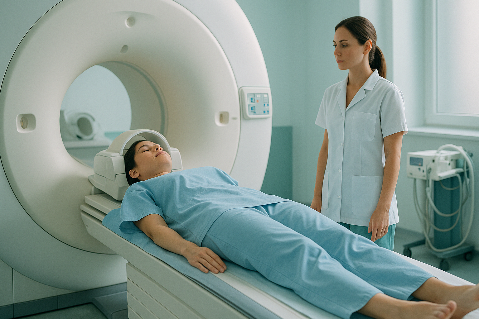
An MRI machine uses a powerful magnet inside a tube-shaped opening, called the bore. The magnet and the receiver coils are built around a flat bed, so you must lie down for the scan to work. This gives excellent detail of soft tissue. But it takes gravity out of the picture. Your spine, pelvis and joints are not carrying their normal standing load when the images are taken.
Key points
- An MRI needs you to lie down because of how the magnet, bore and coils are designed.
- Lying flat takes away gravity, so your body is not carrying its normal standing load.
- Disc height, spinal curves and the spacing of joints can change between lying and standing.
- MRI is the best tool for soft tissue, discs and nerves. A standing X-ray is better for alignment under load.
- The two scans answer different questions, and sometimes you need both.
How does an MRI machine work, and why do you have to lie down?
An MRI machine makes images using a strong magnetic field and radio waves, not radiation. The magnet sits inside a round tube, the bore, which is usually about 60 cm wide. Receiver coils are placed closely around the part of the body being scanned. They pick up faint signals from the hydrogen atoms in your body. For those signals to be clear, you need to stay very still, often for 20 to 40 minutes.
The whole system, the bore, the table and the coils, is built around a horizontal position. You slide into the bore on a flat bed because the magnet's field runs along that line. Building a machine any other way would need a completely different, and very expensive, magnet design.
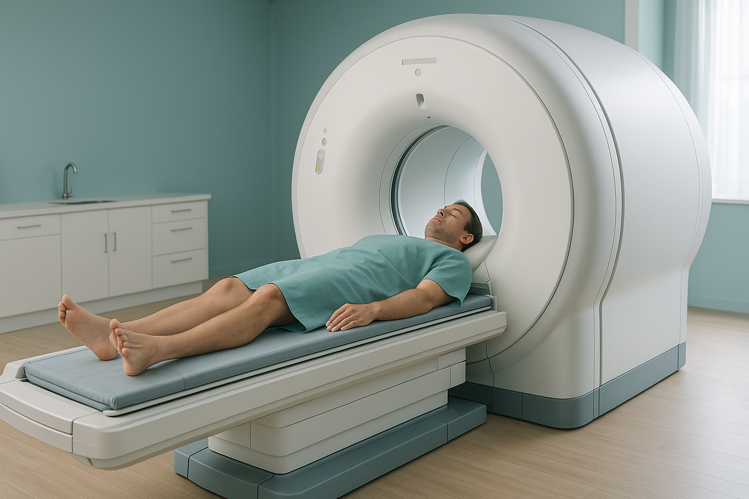
The shape of the bore keeps the magnetic field even and strong across the area being scanned.
The coils must sit close to your body. The flat bed makes positioning exact and easy to repeat.
Lying still is easier than standing still for a long time. This reduces blurring from movement in the images.
What changes in your body when you lie down for a scan?
Quite a lot. When you stand, gravity presses down on your spine and loads your hips and knees. Your muscles work to hold you upright. Lying flat takes all of that away. Your spinal discs expand slightly and your curves flatten. Your pelvis settles into a neutral resting position. This may be quite different from the way it sits when you are on your feet.
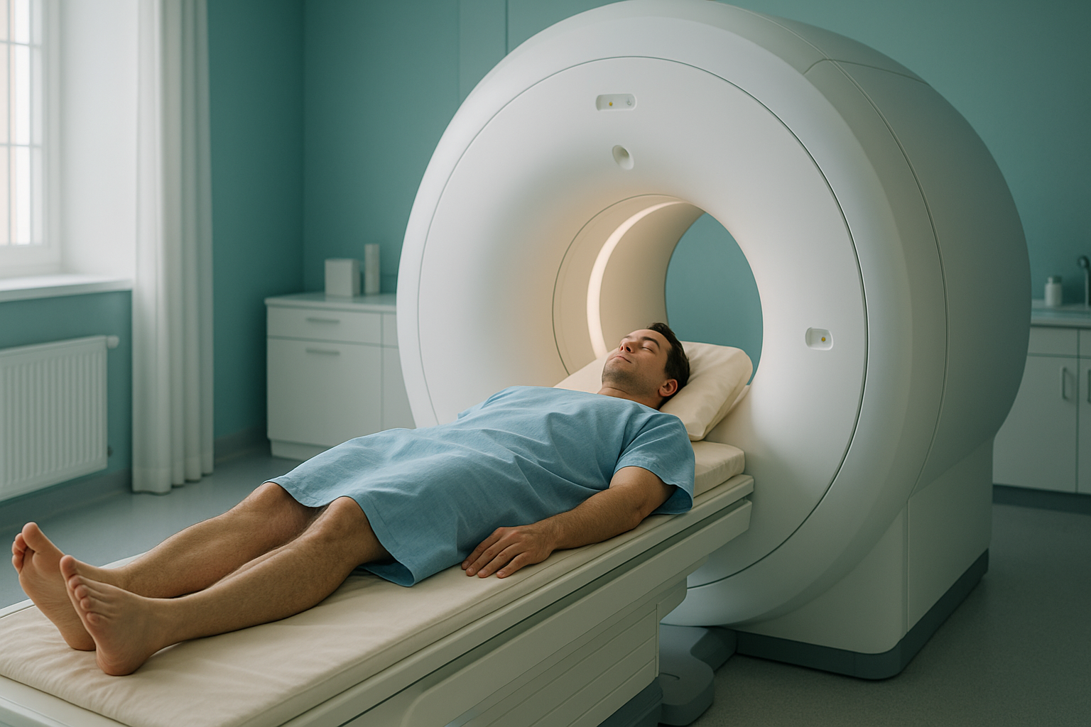
This matters for diagnosis. Research shows that disc height can increase by several percent when you lie down. Spinal curves shift. A tilt or imbalance that causes pain all day may look much milder, or even disappear, on a lying-down scan. Your clinician may be trying to understand your sagittal balance, the overall front-to-back alignment of your skeleton. If so, a lying-down image may not tell the full story.
This is not a fault of MRI. It is simply a limit of the position. MRI was designed to look at structures in fine detail, and it does that brilliantly. But it was not designed to show how your skeleton behaves under the force of gravity. For a fuller comparison, read about weight-bearing versus lying imaging.
Can you have a scan done standing up instead?
Yes, for bone and joint alignment. EOS is a low-dose X-ray system. It scans your whole body from head to feet while you stand naturally. Because you are upright and carrying your own weight, the image shows how gravity, muscle tension and posture all work together in real life.
Your body is a connected chain. Your feet, knees, hips, pelvis and spine all affect one another. For example, a slight twist in the pelvis can change the load on a knee or shift your spinal curves. Pain often appears away from the place causing the problem. A standing whole-body image lets a clinician see the ways your body makes up for this in one view. Lying-down imaging cannot show this.
EOS uses about 90% less radiation than a standard X-ray, around ten times less. You can learn more about how this differs from traditional scans in our guide to weight-bearing or traditional imaging.
You may have been told your MRI looks normal, but you are still in pain. A standing scan may show alignment problems that lying-down imaging cannot capture.
Book a free video consultationWhen is MRI still the right choice?
MRI is still the best imaging tool for many medical questions. Nothing on this page should put you off having one when it is needed.
When you need to see soft tissue, MRI is usually the better tool. EOS and standing X-ray show bones, joints and alignment clearly. For discs, nerves, ligaments, cartilage or any suspected soft-tissue injury, MRI gives the detail that X-ray-based imaging cannot. Many people benefit from both: MRI for structures, and standing imaging for alignment under load.
- Disc bulges or slipped discs
- Pressure on nerves
- Ligament or tendon tears
- Cartilage damage
- Checking the spinal cord
- Whole-body skeletal alignment
- Pelvic tilt or rotation
- Differences in leg length
- Spinal balance under gravity
- Planning before joint surgery
Sometimes the two work together. An MRI may show a disc problem, while a standing scan shows the alignment problem that is overloading that disc in the first place. ScanAlign standing EOS imaging focuses on the alignment side of that picture.
Before you start a treatment, or change the one you are on, it is worth getting a detailed diagnosis. Knowing whether the cause sits in your pelvis, your legs or your spine changes what will actually help.
Frequently asked questions
The machine's magnet sits inside a tube-shaped bore. The receiver coils are designed to wrap around you while you lie on a flat bed. The whole system is built for a horizontal position, to keep the magnetic field even and the images sharp.
Open upright MRI machines do exist. But they are uncommon in the UK and generally give lower-resolution images than standard machines. For bone and joint alignment, a standing EOS X-ray is a more widely available option. It captures your skeleton under normal gravity.
Yes. Without gravity, your discs expand, your spinal curves flatten and your pelvis rests in a neutral position. Alignment problems that cause symptoms under upright load may look milder or not show at all.
EOS is a low-dose X-ray system that scans your whole body while you stand. It uses about 90% less radiation than a standard X-ray, around ten times less. The image shows your bones, joints and alignment from head to feet in a single view.
They answer different questions. MRI is excellent at showing soft tissue such as discs, nerves and ligaments. A standing EOS scan shows how your bones and joints line up under gravity. Neither replaces the other, and many people benefit from both.
No. You can book yourself. The process starts with a free video consultation. A specialist radiographer reviews your history and confirms that a standing scan is suitable for you.
ScanAlign is a private self-pay clinic. The price for patients travelling from abroad is confirmed during the free video consultation, which you can start from the book a standing scan page.
Yes. EOS uses about 90% less radiation than a standard X-ray, around ten times less. It is one of the lowest-dose imaging options available for checking bones and joints.
Pain can come from problems with alignment or how load is spread through the body. These problems only show up under upright load. Because an MRI is done lying down, they may not be visible. A standing whole-body scan can show tilts, rotations or ways your body makes up for them that a lying-down scan misses.
You stand naturally inside the machine for about 20 seconds. Two low-dose X-ray beams capture a front view and a side view. There is no closed tube and no need to hold an awkward position. The whole appointment is quick and simple.
Wondering if a standing scan could help?
A free video consultation with a specialist radiographer will help you understand whether standing imaging is right for your situation, before you plan any travel to London.
Book a free video consultationWritten by Abbas Dhami (Specialist Diagnostic Radiographer)
Sources
- Defined by Design: The Physics of MRI. Radiological Society of North America (RSNA) educational resources.
- NHS guidance: Having an MRI scan. nhs.uk.
- Deschênes S et al. Diagnostic imaging of spinal deformities: Reducing patients' radiation dose with a new slot-scanning X-ray imager. Spine, 2010;35(9):989-994.
This page is general information and not a diagnosis.
ScanAlign is the trading name of CoreMed Solutions Ltd and operates under The Harley Street Hospital's CQC licence.