
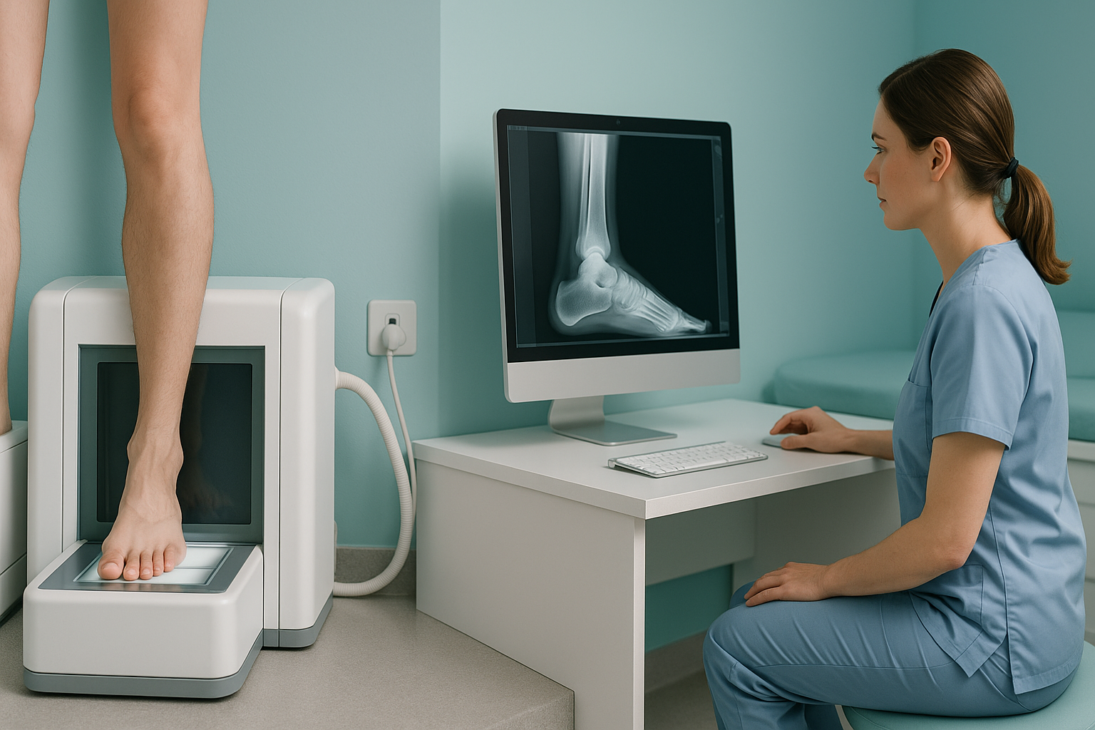
What Is the Evidence Base for Weight-Bearing Imaging?
More and more peer-reviewed research shows that imaging the body while standing reveals changes in alignment, joint space and spinal curves. These are hidden when a patient lies down. Studies consistently find clinically significant differences between supine (lying) and standing measurements. This makes weight-bearing imaging a truer picture of how your skeleton actually works.
Key points
- Standing X-rays often show larger spinal curves and narrower joint spaces than lying-down images of the same patient [1, 2].
- Biplanar EOS imaging uses about 90% less radiation than a standard X-ray, around ten times less, while producing full-length, weight-bearing images [3].
- Research supports more reliable measurements (lower intra-observer variation) with calibrated standing systems than with conventional films.
- Weight-bearing imaging has known limitations. It shows bones and alignment, not soft tissue, and most studies use moderate sample sizes.
- For soft-tissue, disc or nerve problems, MRI remains the better tool.
If you have an ongoing back, hip or knee problem, you may already have had scans that came back "normal". One reason for this is that many conventional scans are taken lying down. The evidence on this page explains why that matters, what the research actually shows, and where the evidence currently falls short. For a plain-language comparison, see the guide to weight-bearing versus lying imaging.
Why does position matter when you have an X-ray?
Position matters because gravity changes the shape and loading of your skeleton. When you lie flat on a table, your muscles relax, your spine straightens slightly and your joints decompress. So an image taken supine (lying down) may not reflect what is happening when you stand, walk or sit, the positions in which you actually feel pain.
Many studies have recorded measurable differences between supine and standing X-rays, in both the spine and the legs [1, 2]. These are not small academic points. A curve that looks mild lying down may cross a clinically relevant threshold when the patient stands. A knee joint that looks well spaced lying down may show significant narrowing under load.
What does the research say about standing spine imaging?
Research consistently shows that spinal curves are larger when measured standing than when measured lying down. A systematic review in the European Spine Journal found that sagittal parameters, the natural front-to-back curves of the spine, change significantly between supine MRI and standing X-rays [1]. This matters directly for care, because decisions about surgery and bracing are based on these measurements.
Sagittal balance, the way the spine's curves work together to keep your head centred over your pelvis, is a good example. Sagittal balance can only be assessed properly when the patient is upright and bearing weight. An MRI or CT taken lying down simply cannot capture it.
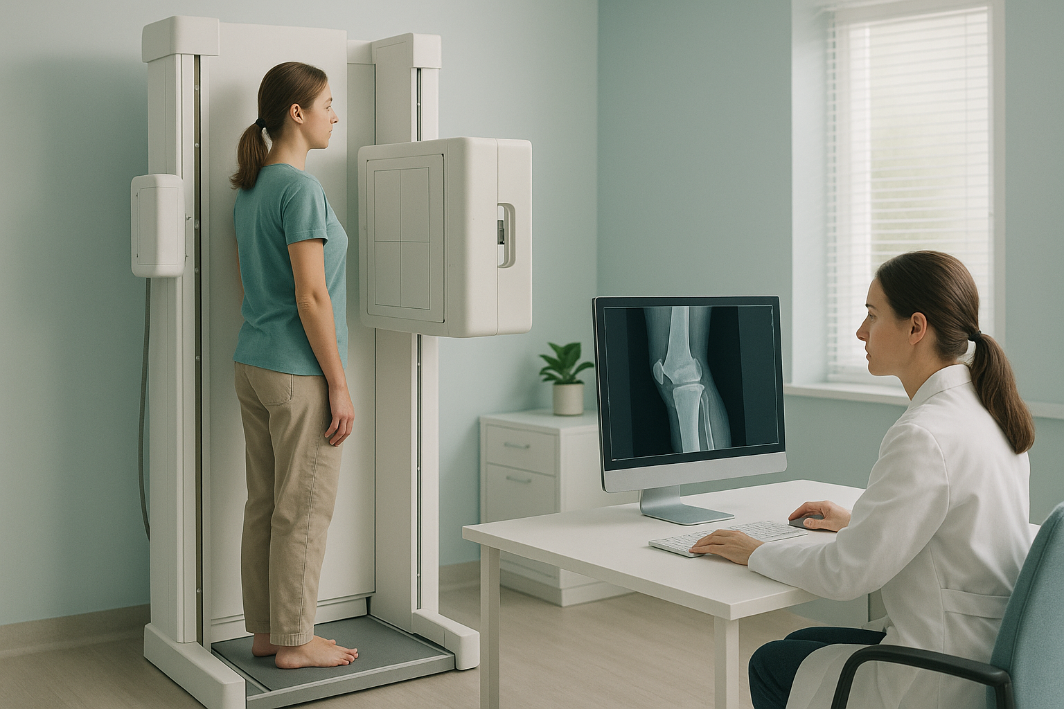
Biplanar EOS systems address this. They produce a calibrated, full-length standing image of the spine, from head to pelvis, in a single exposure. Published studies report high intra-observer and inter-observer reliability for spine measurements made from EOS images. This means different radiologists measuring the same image tend to reach very similar numbers [3].
What it shows well: Detailed cross-sectional anatomy of discs, nerves and soft tissue (MRI). Bone detail in fine slices (CT).
What it may miss: True spinal curves under gravity. Sagittal balance. Compensatory patterns between regions. The way nearby joints take load under body weight.
What it shows well: Real-life alignment under gravity. Full-length skeletal shape from head to feet. Joint spacing as it is when you stand and walk.
What it may miss: Soft-tissue detail (discs, ligaments, muscles). Internal organ structures. Subtle bone-marrow changes.
Is there evidence for weight-bearing imaging of the knee and hip?
Yes. Research in lower-limb orthopaedics has long recognised that weight-bearing X-rays show joint-space narrowing more accurately than lying-down images. A study in Knee Surgery, Sports Traumatology, Arthroscopy found that standing knee X-rays showed greater and more clinically representative joint-space loss than lying-down films of the same patients [2]. This affects how arthritis is graded and whether surgery is recommended.
For leg alignment, a full-length standing image captures the mechanical axis of the whole leg, from hip to ankle. Surgeons planning knee or hip replacements increasingly use this information to guide where the implant goes. Conventional short-film X-rays cannot show the whole mechanical axis in one exposure. This limits their use for planning before surgery.
| Research area | Standing finding | Clinical relevance |
|---|---|---|
| Knee joint space | Narrower under load than lying down | More accurate arthritis grading |
| Mechanical axis (whole leg) | Visible only on full-length standing film | Better surgical planning |
| Spinal sagittal parameters | Curves larger standing than lying down | Truer assessment of deformity |
| Pelvic tilt | Changes between lying and standing | Affects how hip and spine interact |
How accurate is EOS imaging and what is the radiation dose?
EOS imaging uses about 90% less radiation than a standard X-ray, around ten times less. This low dose is well documented in the manufacturer's published data and in independent dosimetry studies [3]. It matters because full-length imaging from head to foot would involve a significant dose on a conventional system, especially if several views are needed.
Accuracy studies have looked at both 2D measurements (distances and angles on the flat image) and 3D reconstructions made by the EOS software. Published research reports high measurement reliability, with low intra-observer variation. This means the same clinician measuring the same image on different occasions gets consistent results [3]. Inter-observer reliability (agreement between different clinicians) is also reported as strong. The exact figures vary by study and by which parameter is measured.
It is worth noting that most of these studies use moderate sample sizes, usually dozens to low hundreds of patients. Large randomised controlled trials comparing clinical outcomes (not just measurement accuracy) between standing and lying-down imaging are still fairly few. The evidence supports standing imaging as more representative, but the field would benefit from larger outcome studies.
What are the limitations of the current evidence?
An honest look at the evidence is important. Here are the main limitations to be aware of.
Many studies involve moderate groups of patients. Larger trials across several centres would strengthen the evidence further.
Most studies show that standing measurements are different and more reliable. Fewer studies show that this leads to better clinical outcomes, such as better surgical results.
Weight-bearing X-ray systems, including EOS, show bones and joints. They do not image discs, nerves, ligaments or muscles. MRI is needed for these.
A patient who cannot stand without help, or who has significant pain standing still, may not produce an ideal weight-bearing image.
None of these limitations cancels out the main finding. They simply mean the evidence is still developing, and clinicians should read results alongside the full clinical picture.
A note on MRI. EOS and other standing X-ray systems show bones, joints and alignment very clearly. For soft-tissue problems, such as disc herniations, nerve compression, ligament tears or muscle injuries, MRI is usually the better tool. The two types of imaging answer different questions, and the best assessment often uses both. If you want to know whether weight-bearing or traditional imaging is the right place to start for you, a short consultation can help.
Before you start a treatment, or change the one you are on, it is worth getting a detailed diagnosis. Knowing whether the cause sits in your pelvis, your legs or your spine changes what will actually help.
If you are a clinician who wants to refer a patient, or you would like to know whether standing imaging suits your situation, you can discuss a referral with our team at no cost.
Book a free video consultationFrequently asked questions
Yes. Many peer-reviewed studies and systematic reviews confirm that standing images reveal alignment differences and joint-space changes that lying-down imaging misses [1, 2, 3]. The evidence is growing, though larger outcome trials are still needed.
EOS uses about 90% less radiation than a standard X-ray, around ten times less. This is documented in published dosimetry studies and makes full-length imaging practical with a very low dose.
Yes. Research consistently shows measurable differences. Spinal curves tend to be larger standing, and knee joint spaces tend to be narrower under load. These differences can change clinical decisions.
No. EOS shows bones, joints and skeletal alignment. MRI shows soft tissue such as discs, nerves and ligaments. They answer different questions. Many patients benefit from both.
Published studies report high intra-observer and inter-observer reliability for EOS measurements. This means different clinicians measuring the same image reach similar results. Reliability varies slightly depending on which parameter is measured.
EOS is uncommon in the UK but is available at a small number of private clinics. ScanAlign, standing EOS imaging, is based at 19 Harley Street, London, and offers self-referred appointments.
No. You can refer yourself. The process starts with a free video consultation, where a specialist radiographer reviews your history and confirms whether a standing scan is appropriate.
Most studies have moderate sample sizes and focus on measurement differences rather than long-term clinical outcomes. Patients who cannot stand without help may not be suitable. Soft-tissue problems need MRI.
One common reason is that conventional scans taken lying down can hide alignment problems that only appear under gravity. A standing image shows your skeleton as it actually works, which may reveal a cause your previous scan did not capture.
For patients travelling from abroad, the price is confirmed during the free video consultation, which carries no obligation.
See what standing imaging could show
Start with a free, no-obligation video consultation. A specialist radiographer will review your history and advise whether a standing scan is appropriate.
Book a free video consultationSources
- Le Huec, J.C., et al. "Sagittal balance of the spine." European Spine Journal, 2019. A systematic review of differences in sagittal parameters between lying-down and standing imaging.
- Brouwer, R.W., et al. "Association between valgus and varus alignment and the development and progression of radiographic osteoarthritis of the knee." Arthritis & Rheumatism, 2007. Shows the clinical importance of assessing leg alignment while bearing weight.
- Illés, T. and Somoskeöy, S. "The EOS imaging system and its uses in daily orthopaedic practice." International Orthopaedics, 2012. A review of EOS dosimetry, measurement reliability and clinical uses.
Written by Abbas Dhami (Specialist Diagnostic Radiographer).
This page is general information and not a diagnosis.
ScanAlign is the trading name of CoreMed Solutions Ltd and operates under The Harley Street Hospital's CQC licence.