
What Is Tibial Torsion, and How Is It Assessed?
Tibial torsion is a twist along the length of the shin bone (tibia). Everyone has some rotation in the tibia. But when there is too much, or too little, it can change the way you walk, put stress on your knees, and cause pain in places you might not expect. To measure it accurately, the scan needs to capture your legs while you are standing and bearing weight.
Key points
- Tibial torsion means your shin bone is twisted inward (internal) or outward (external) more than the typical range.
- It can cause in-toeing, out-toeing, knee pain and uneven shoe wear.
- Lying-down scans relax the muscles and can hide the true rotation. Standing imaging captures your real, weight-bearing alignment.
- Rotation in the tibia often comes together with rotation in the hip. Both need to be measured together as part of a full lower limb alignment assessment.
- EOS whole-body imaging uses about 90% less radiation than a standard X-ray, around ten times less.
Your body is a connected chain from your feet up to your head. The tibia sits right in the middle of that chain. If it is twisted beyond the normal range, your kneecap, ankle and foot all shift their position. Over time, nearby joints compensate. Symptoms can then appear far from the actual problem.
What are the signs of tibial torsion in adults?
The most common signs are changes in the way your feet point when you stand or walk. You may also notice aching around the knee with no obvious injury behind it.
When you stand relaxed, your feet naturally point inward or outward more than expected.
A twisted tibia changes how your kneecap tracks. This can lead to persistent anterior knee pain.
Shoes wear down much faster on one side, or differently between the left and right foot.
When the feet point in, they can catch on each other, especially when you are tired or walking quickly.
What causes tibial torsion?
Tibial torsion develops for several reasons. Some people are simply born with more or less twist in the shin bone. In others, an old fracture, surgery or a long-standing abnormal loading pattern has changed the bone's rotation over time.
In some adults, the natural twist that forms as the bone grows settles outside the typical range.
A healed tibial fracture can set with a rotational shift. This changes the bone's direction permanently.
A knee replacement or tibial osteotomy can change rotation if the component is placed with a slight twist.
Importantly, tibial torsion rarely exists alone. The hip and femur often rotate the opposite way to compensate. This pattern is known as anteversion versus retroversion. That is why measuring one bone on its own only tells part of the story.
What is the difference between internal and external tibial torsion?
The direction of the twist decides the type. Internal tibial torsion means the shin bone is rotated inward compared with the thigh bone above it. External tibial torsion means it is rotated outward. Both can cause problems, but they load the knee and ankle in different ways.
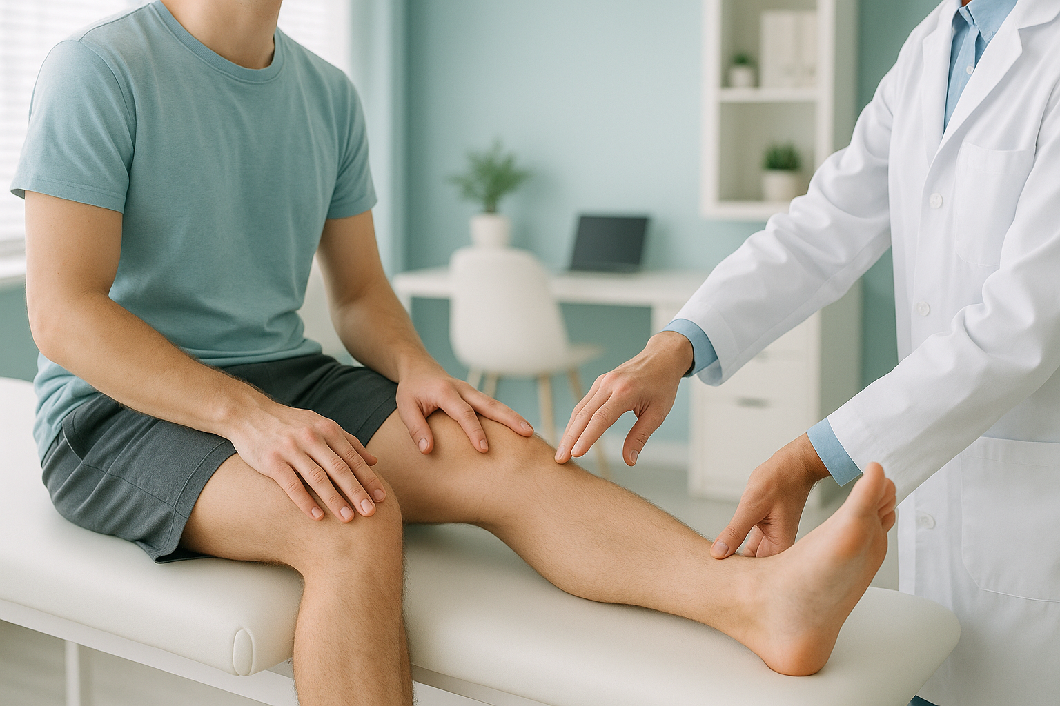
Internal tibial torsion turns the foot inward (in-toeing). The kneecap tends to face forward while the ankle and foot angle inward. This puts extra stress on the outer side of the knee. It can be linked to IT band tightness and lateral compartment overload.
External tibial torsion turns the foot outward (out-toeing). This is more common in adults. It is linked with more stress on the inner (medial) side of the knee. It can contribute to anterior knee pain, patellar maltracking and a flat-footed gait.
How is tibial torsion measured?
Tibial torsion is measured by comparing the direction of the knee joint line with the direction of the ankle joint line. Clinicians sometimes call this the transmalleolar angle or bimalleolar axis. There are several methods.
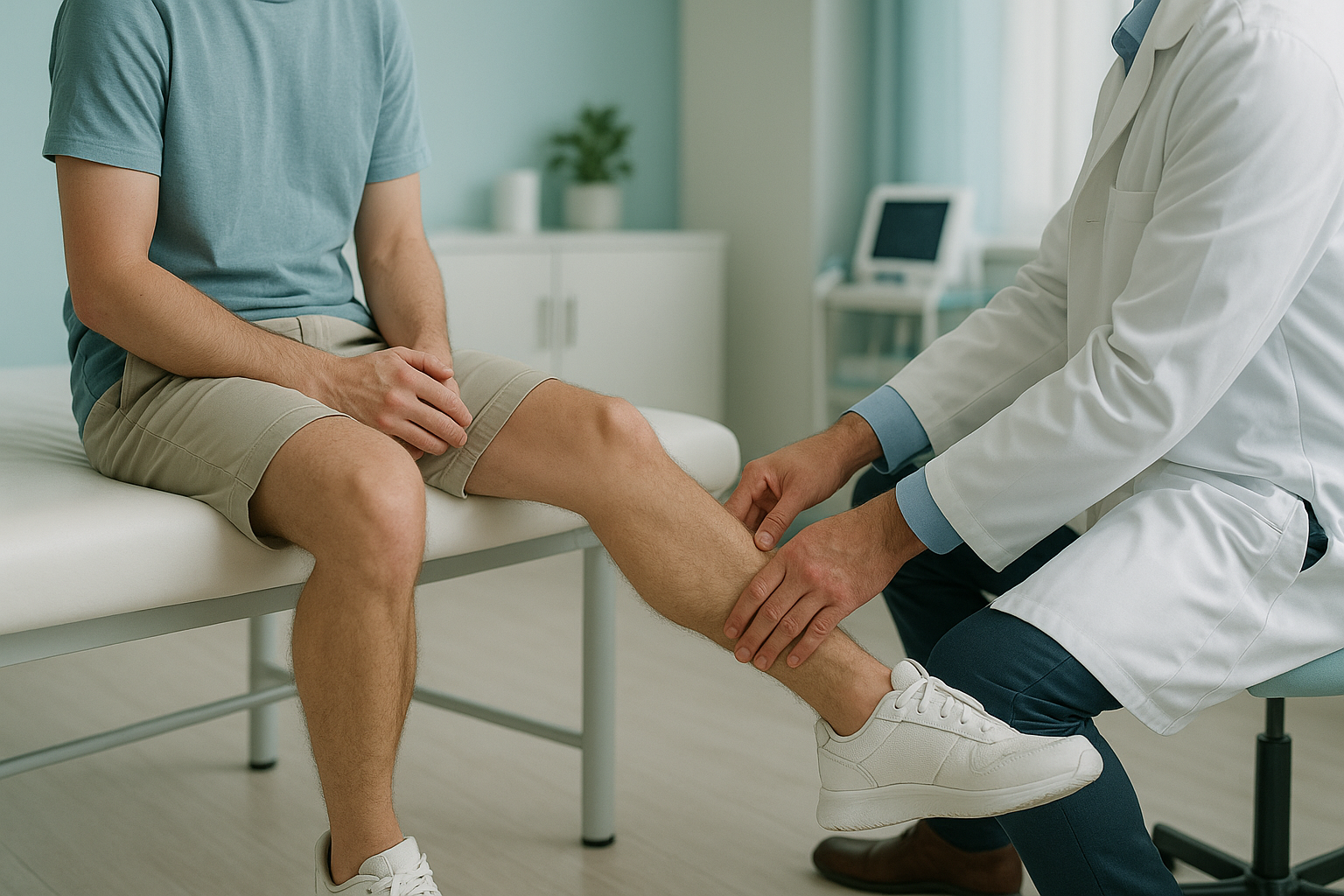
- Clinical examination. A clinician estimates the thigh-foot angle while you lie face-down with your knee bent. This is a useful screening step. But it depends on the examiner's judgement and cannot give a precise rotational profile.
- CT rotational study. A CT scan takes cross-sectional slices at the knee and the ankle to measure the twist. It is accurate, but you lie down for it. This removes the effect of gravity and muscle activity on alignment. It also involves higher radiation.
- EOS standing imaging with 3D reconstruction. An EOS scan captures your entire lower limb, head to foot, while you are standing. Software then builds a 3D model of each bone. This allows the exact rotational profile to be measured under real, weight-bearing conditions.
Rotation in the tibia is often part of a larger pattern that includes the hip and femur. So measuring only the shin bone on its own can miss the bigger picture. A full rotational profile, from pelvis to ankle, is more clinically useful.
Why does a standing scan matter for shin bone rotation?
When you lie down for a CT or a standard X-ray, your muscles relax and your joints settle into a resting position. Rotational misalignments that cause real problems under load can look less severe in this relaxed state, or even disappear.
Standing imaging captures how your bones, joints and muscles actually behave under your body weight. This matters a lot for tibial torsion. Your foot position, knee tracking and compensating hip rotation all change the moment gravity acts on them.
At ScanAlign, 3D standing skeletal imaging, the EOS system captures a full-length image of your skeleton in a single, low-dose scan. It uses about 90% less radiation than a standard X-ray, around ten times less. The 3D reconstruction then allows precise rotation measurements at every level. This gives your clinician a complete rotational profile rather than separate snapshots.
Would you like to understand your lower limb rotation under real, weight-bearing conditions?
Book a free video consultationWhat can be done about tibial torsion?
Management depends on how much torsion you have, whether it is causing symptoms, and whether other parts of your alignment are compensating. ScanAlign is a diagnostic imaging clinic, so we do not provide treatment. But an accurate measurement of your rotational profile is the essential first step for any specialist who may be advising you.
In general, options your treating clinician may consider include:
- Physiotherapy and gait retraining to strengthen the muscles that control rotation and improve movement patterns.
- Orthotics to adjust foot position and reduce compensating stress on the knee.
- Surgical correction (derotation osteotomy) where the torsion is significant and causing symptoms. This involves carefully cutting and re-aligning the bone.
All of these decisions depend on starting from precise, weight-bearing measurements. That is the value of accurate imaging.
Before you start treatment, or change the one you are on, it is worth getting a detailed diagnosis. Knowing whether the cause sits in the pelvis, the legs or the spine changes what will actually help.
When is MRI more useful? EOS imaging shows bones, joints and alignment. If your clinician suspects a soft-tissue problem such as cartilage damage, ligament injury or nerve involvement, MRI is usually the better tool. Sometimes both are needed to build a complete picture.
- Have in-toeing or out-toeing with no clear explanation
- Have persistent knee pain without a clear cause
- Had a tibial fracture and now walk differently
- Are considering corrective surgery and need a baseline
- Were told your alignment is fine on a lying-down scan but still have symptoms
- Have sudden swelling, locking or giving way (you may need an MRI first)
- Have numbness, weakness, or loss of bladder or bowel control. Please get urgent medical help from your doctor or the nearest emergency department
- Have a soft-tissue injury that needs detailed views of the ligaments or cartilage
Frequently asked questions about tibial torsion
Yes. Tibial torsion is the medical term for a shin bone that is rotated along its length more than the typical range. "Twisted shin bone" is a perfectly accurate way to describe it.
Yes. A twisted tibia changes the way your kneecap tracks and alters the forces through the knee joint. This can lead to anterior knee pain, patellar maltracking and more wear on one side of the knee.
Tibial torsion is rotation in the shin bone (tibia). Femoral torsion is rotation in the thigh bone (femur). They often exist together. When the femur twists one way, the tibia may compensate by twisting the other way. A full rotational profile measures both. You can learn more about the femoral side on our page about anteversion versus retroversion.
No. ScanAlign accepts self-referrals. You can book a rotation measurement scan directly, or start with a free video consultation to discuss whether this imaging is right for you.
The rotation is measured by comparing the alignment of the knee joint line with the ankle joint line. On EOS imaging, a 3D reconstruction of the entire lower limb allows this to be calculated precisely while you are standing and bearing weight.
Yes. EOS uses about 90% less radiation than a standard X-ray, around ten times less. It is a very low-dose system designed for full-body imaging.
In many cases, physiotherapy, gait retraining and orthotics can manage symptoms effectively without surgery. Treatment decisions depend on the severity and the symptoms. Accurate imaging gives your treating clinician the measurements they need to advise you.
For patients travelling from abroad, the price is confirmed during the free video consultation.
When you lie down, gravity no longer acts on your joints and your muscles relax. Rotational problems that cause symptoms when you stand and walk can settle into a more neutral position on the table. The scan can then look normal when it may not be.
Absolutely. It is common for the left and right tibia to have different amounts of rotation. That is one reason a full standing scan of both legs is important. It allows your clinician to compare both sides under the same weight-bearing conditions.
Understand your rotation, standing up
A free video consultation lets you discuss your symptoms with our specialist radiographer from home, before you plan any travel to London. You can find out if a standing rotation scan is the right next step.
Book a free video consultationWritten by Abbas Dhami (Specialist Diagnostic Radiographer)
Sources
- Staheli LT. Rotational problems in children. Journal of Bone and Joint Surgery, 1993;75-A(6):939-949.
- Guenoun B, et al. Reliability of a new method for lower-extremity measurements based on stereoradiographic three-dimensional reconstruction. Orthopaedics & Traumatology: Surgery & Research, 2012;98(5):506-513.
- Royal College of Radiologists. iRefer Guidelines: Making the Best Use of Clinical Radiology. RCR, London (current edition).
This page is general information and not a diagnosis.
ScanAlign is the trading name of CoreMed Solutions Ltd and operates under The Harley Street Hospital's CQC licence.