
How Do Scoliosis Therapists Use Serial Imaging?
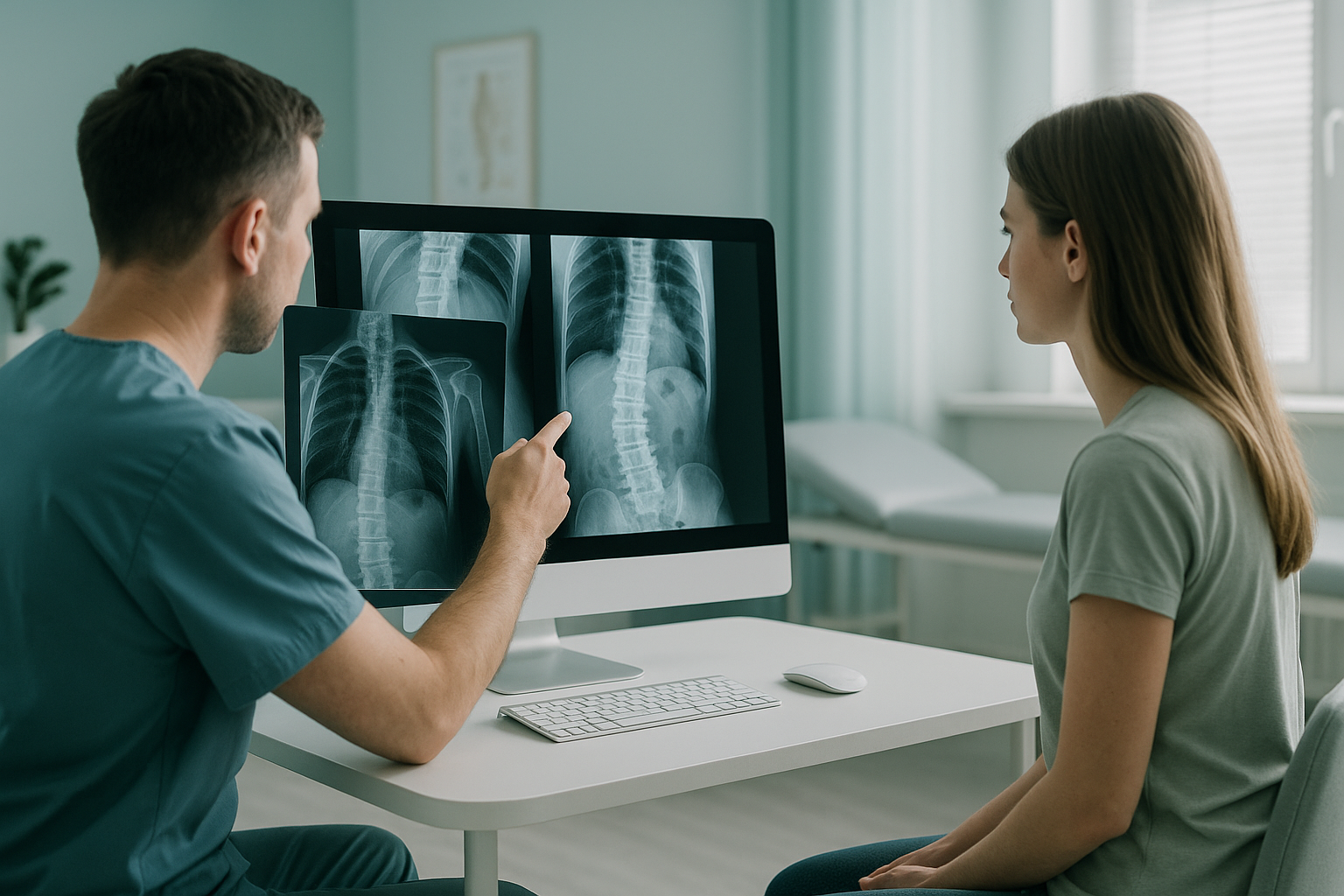
Scoliosis therapists use serial imaging to compare standing whole-body scans taken at intervals over time. They measure the same landmarks on each scan. This shows whether a curve is stable, improving or progressing. This objective evidence helps guide exercise programmes, brace adjustments and decisions about onward referral.
Key takeaways
- Serial imaging gives therapists a consistent record of how a curve behaves over months or years, with each scan comparable to the last.
- Standing scans capture alignment under real weight-bearing load, which lying-down imaging can miss.
- Low-dose EOS imaging uses about 90% less radiation than a standard X-ray, around ten times less, making repeat scans more practical.
- Standardised Cobb angle measurement on every scan reduces variation between the people reading the scans.
- Therapists can register to refer patients directly for imaging without a letter from the patient's doctor.
Why do scoliosis therapists need serial imaging?
Serial imaging gives therapists objective, dated evidence of how a curve is responding to treatment. Without it, judging outcomes relies on clinical observation alone. That is valuable, but it cannot reliably pick up small changes in the size of a curve.
You may deliver Schroth-based exercise, SEAS or any other scoliosis-specific programme. Either way, your clinical reasoning depends on knowing whether a curve is stable, reducing or progressing. A single baseline scan tells you where things stand today. Follow-up scans over time tell you whether your treatment is working.
How is curve change measured reliably between scans?
Curve change is measured by comparing the same anatomical landmarks on standing images taken one after another. The key measure is the Cobb angle, taken between the most tilted vertebrae above and below the curve apex. When the same imaging system, positioning protocol and measurement method are used each time, variation between readers drops. Real change then becomes easier to tell apart from measurement noise.
Research suggests that the built-in error for manual Cobb angle readings on plain film X-rays can be several degrees. A consistent digital platform and a standard way of choosing the end vertebrae narrow that margin. That is why imaging on the same system matters. You can read more about how the measurement works on our Cobb angle measurement page.

For therapists, the practical point is simple. To know whether a curve has really changed, you need at least two comparable images taken the same way. When you request imaging through ScanAlign, low dose standing imaging, the positioning protocol is the same every time. This makes your serial comparisons as reliable as possible.
Is serial imaging safe when patients need repeated scans?
Total radiation exposure over time is a fair concern whenever repeated imaging is planned. EOS technology uses about 90% less radiation than a standard X-ray, around ten times less. That lower dose per scan makes it far more practical to build a useful series of images over months or years without excessive total exposure.
For therapists managing adults with scoliosis, this changes the conversation. You no longer need to ration imaging because of dose concerns. You can request a follow-up scan when it is clinically useful, whether at six months, twelve months or any interval your clinical judgement calls for.
If you are a scoliosis therapist and would like to refer patients directly for standing imaging in London, you can register to refer patients through our online form.
Why does standing imaging matter more than lying-down scans?
Standing imaging captures the spine under real gravitational load. This is how your patient lives, moves and feels their symptoms. When a patient lies down for a conventional X-ray, CT or MRI, gravity is removed and muscles relax. The curve can then look different from how it truly behaves in daily life.
What it shows: The full weight-bearing alignment of the spine, pelvis, hips and legs in one image. Compensatory shifts, trunk offset and pelvic obliquity are visible as they actually occur in daily life.
Why it matters for therapists: You see the alignment your exercises are trying to change, not a relaxed version of it.
What it shows: Spinal anatomy without gravitational load. Useful for assessing soft tissue and discs, but curves can look smaller or different when the patient is lying on their back.
The gap for therapists: A lying-down image may underestimate the curve your patient actually lives with. This can mislead outcome measurement.
This difference is especially important for serial monitoring. If one scan is taken standing and the next lying down, any apparent change could be due to position rather than real curve behaviour. Consistency matters.
How do therapists refer a patient for serial imaging?
The referral process is designed to be simple, so you can focus on clinical care rather than paperwork.
Fill in a short online form to register to refer patients. Once approved, you can send referrals whenever you need to.
When a patient needs imaging, refer a patient through the booking system. Include clinical details so the radiographer can tailor the protocol.
Your patient comes to 19 Harley Street, London, for a standing whole-body EOS scan. No special preparation is needed. The scan itself takes only a few seconds.
The report, including relevant measurements, is shared with you and the patient. You can compare it directly with earlier scans.
When should a therapist request MRI instead?
EOS is excellent at showing bone alignment, how joints relate to each other and overall posture under load. For assessing soft tissue, discs, the spinal cord or nerve roots, MRI is usually the better tool. If your patient has neurological symptoms or radiculopathy, or you suspect a disc is involved, an MRI referral is more suitable for that specific question.
A note on MRI: EOS shows bones, joints and alignment. For soft tissue, discs or nerves, MRI is usually the better tool. Many patients benefit from both, as each answers a different clinical question.
- Tracking curve size over time
- Measuring pelvic obliquity and trunk shift
- Comparing in-brace and out-of-brace alignment
- Assessment before and after an exercise programme
- Whole-body alignment from head to feet
- You suspect a disc herniation or nerve compression
- Neurological signs are present
- The spinal cord needs to be assessed
- Soft-tissue detail is the main question
Before starting treatment, or changing the one a patient is already on, it is worth getting a detailed diagnosis. Knowing whether the cause sits in the pelvis, the legs or the spine changes what will actually help.
Common questions from scoliosis therapists
No. Qualified scoliosis therapists, physiotherapists, osteopaths and chiropractors can refer patients directly. You register once through our online referral form. You can then send referrals as needed.
There is no single rule. Many therapists request a baseline scan and then follow up at six or twelve months. The interval depends on your clinical judgement and the patient's situation. The low dose makes repeated imaging more practical.
EOS uses about 90% less radiation than a standard X-ray, around ten times less. This is particularly relevant for serial monitoring, where the total dose over time matters.
You can compare measurements such as the Cobb angle. But differences in positioning and imaging method may introduce variation. For the most reliable serial comparison, using the same system and protocol each time is ideal.
The report includes a radiologist's assessment of the images and relevant alignment measurements. You receive it in a format you can use alongside your clinical notes and share with the patient's wider care team.
Yes. In-brace and out-of-brace scans can be taken during the same visit. This gives you a direct comparison of the brace's effect under identical conditions.
The price for patients travelling from abroad is confirmed during the free video consultation. ScanAlign is a private, self-pay service, so patients pay directly without needing insurance authorisation.
Yes. Standing whole-body imaging is well suited to adult scoliosis, whether idiopathic or degenerative. It captures the full alignment picture, including sagittal balance, which is often the more important plane in adult curves.
Yes, of course. You can book a free video consultation to discuss whether imaging is suitable and which protocol would best answer your clinical question.
ScanAlign is based at 19 Harley Street, London, within The Harley Street Hospital. The location can be reached by public transport and has step-free access to the imaging suite.
Ready to refer a patient?
Register once, then send referrals whenever your patients need standing imaging. Or discuss a case with us first.
Book a free video consultationSources
- Illés T, Somoskeöy S. The EOS imaging system and its uses in daily orthopaedic practice. International Orthopaedics (SICOT). 2012;36(7):1325–1331.
- Society on Scoliosis Orthopaedic and Rehabilitation Treatment (SOSORT). 2016 guidelines on the treatment of idiopathic scoliosis. Scoliosis and Spinal Disorders. 2018;13:3.
- Langensiepen S, Semler O, Sobottke R, et al. Measuring procedures to determine the Cobb angle in idiopathic scoliosis: a systematic review. European Spine Journal. 2013;22(11):2360–2371.
Written by Abbas Dhami (Specialist Diagnostic Radiographer)
This page is general information and not a diagnosis.
ScanAlign is the trading name of CoreMed Solutions Ltd and operates under The Harley Street Hospital's CQC licence.