

What Does Your Sagittal Vertical Axis (SVA) Measurement Mean?
The sagittal vertical axis is a distance. Picture a straight line dropped down from the C7 vertebra (the bony bump at the base of your neck). The SVA is how far that line sits from the back of your sacrum (the base of your spine). It tells clinicians how far forward or backward your upper body leans when you stand. A larger forward measurement often signals that your spine is out of sagittal balance.
Key points
- SVA measures how far forward your head and trunk sit compared with your pelvis when you stand.
- In most adults, a small forward offset is considered normal.
- A larger SVA is linked to back pain, tiredness and difficulty standing upright.
- It can only be measured accurately while you are standing and bearing your own weight.
- EOS imaging captures the full spine in one standing image. It gives a precise SVA reading with about 90% less radiation than a standard X-ray.
What is the sagittal vertical axis?
The sagittal vertical axis (SVA) is a single measurement. It describes the front-to-back alignment of your spine when seen from the side. To take it, a vertical plumb line is drawn down from the centre of the C7 vertebra. Then the distance is measured between that line and the back corner of the sacrum, in front or behind.
Think of it as a simple forward-lean test for your skeleton. If the line falls well in front of your sacrum, your trunk is tipped forward. Clinicians call this "positive sagittal balance." If the line falls behind, that is "negative sagittal balance." A small amount of forward offset is entirely normal.
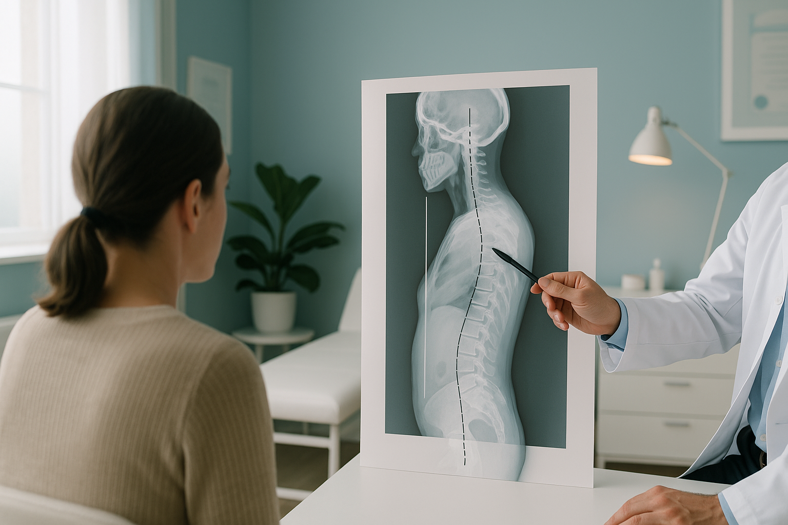
SVA is one of the most widely used measurements in spine assessment. Surgeons, physiotherapists and radiologists rely on it. It helps them see whether your spine can keep your head and trunk centred over your pelvis without too much effort.
What is a normal SVA value?
In healthy adults, the C7 plumb line usually falls within about five centimetres in front of the sacrum. Glassman et al. published research in the journal Spine in 2005. They found that health-related quality-of-life scores drop noticeably when the SVA goes beyond roughly five centimetres of forward offset. The Scoliosis Research Society uses a similar limit in its guidelines.
It is important to know that SVA increases naturally with age. Spinal discs lose height and the mid-back curves a little more. So over the decades, many people develop a slightly larger forward offset. A value that would be unusual at 30 may be quite typical at 70.
This is why your SVA should never be read on its own. It sits alongside other measurements of sagittal balance, including pelvic tilt and the shape of the lumbar and thoracic curves. Together, these numbers give a full picture of how your spine is coping.
What does a high SVA feel like?
A high SVA means your trunk is tipped further forward than expected. You may not notice it at first, because your body adapts. Over time, however, common signs include the following.
Your muscles work harder to hold you upright. This leads to tiredness after short spells of standing or walking.
You may find yourself looking at the ground more often, because your head has moved forward.
Pain often appears in areas that are making up for the forward lean, not necessarily where the problem starts.
You may bend your knees without realising it, to move your weight backward and keep your balance.
Sudden numbness, weakness, or any loss of bladder or bowel control are red-flag symptoms. If you have any of them, please get urgent medical help from your doctor or the nearest emergency department rather than booking a scan.
How does your body compensate for a forward-leaning spine?
Your body is remarkably good at keeping your head over your feet, even when your spine curves further forward than it should. It does this through a chain of compensations that run from head to toe.
Your pelvis tilts backward to bring your centre of gravity closer to your feet.
Bending the knees slightly moves the weight line backward to balance the forward trunk.
The hips straighten, or even overextend, to push the pelvis forward and reduce the offset.
Each of these adaptations uses extra energy and can cause pain well away from the spine. That is why someone with a spinal alignment problem may feel it most in their hips, knees or even their feet. Conditions like flatback syndrome and forward leaning posture are closely linked to a raised SVA. The compensation patterns they cause are often what lead people to seek help.
The key point is that looking at one area alone can miss the full story. A scan of your lower spine might look normal, while the real issue is the overall alignment of the whole chain.
Why does SVA need to be measured standing up?
SVA is a weight-bearing measurement. It describes what your spine does when gravity is pulling on it and your muscles are working to hold you upright. Lying down for a regular X-ray, CT or MRI relaxes those muscles and lets the spine flatten out. This can change the reading a great deal.
Gravity is active. Your muscles, joints and discs carry load exactly as they do in daily life. The SVA reading shows your real-world alignment, including any compensations your body is making.
A full-body standing image captures everything from your skull to your ankles in a single exposure. So the C7 plumb line, the position of the pelvis and the alignment of the legs can all be measured together. This gives a true picture of your sagittal balance.
Gravity is removed. Your spine straightens, curves flatten and muscles switch off. The SVA from a lying-down scan can look much smaller than it really is. This could hide a significant alignment problem.
This does not make lying-down scans useless. They are still the best choice for certain tasks, especially looking at individual discs, nerves or soft tissue. But for alignment measurements like SVA, standing imaging is the clinical standard.
At ScanAlign, unstitched full-body standing imaging uses EOS technology. It captures your entire skeleton in a single standing exposure with about 90% less radiation than a standard X-ray, around ten times less. The image is not stitched together from smaller frames. So measurements like SVA are precise and free from stitching error.
A note on MRI. EOS shows bones, joints and alignment. For soft-tissue problems such as disc bulges, nerve compression or ligament damage, MRI is usually the better tool. Your clinician can advise which scan, or combination, is right for you.
Before you start treatment, or change the one you are on, it is worth getting a detailed diagnosis. Knowing whether the cause lies in your pelvis, your legs or your spine changes what will actually help.
Would you like to understand your spinal alignment with a precise, standing measurement? If you are thinking of travelling to London, you can book a standing spine scan or start with a free video consultation to talk about the imaging you need.
Book a free video consultationFrequently asked questions about SVA
SVA stands for sagittal vertical axis. It is the horizontal distance between a vertical line dropped from the C7 vertebra and the back corner of the sacrum. It is measured while you stand upright.
In most healthy adults, the C7 plumb line falls within roughly five centimetres in front of the sacrum. A small forward offset is normal. Values increase naturally with age, so what counts as normal depends partly on how old you are.
Not necessarily. A mildly positive SVA is common and may cause no symptoms at all. It becomes significant when the forward offset is large enough to cause pain, tiredness or difficulty standing upright. It also matters when your body is relying heavily on compensation to stay balanced.
A standing full-spine X-ray taken from the side can measure SVA. But standard X-rays of a single area usually do not capture enough of the spine. Full-body standing imaging, such as EOS, captures the whole skeleton in one frame. This avoids the stitching errors that happen when several smaller images are joined together.
Lying down removes gravity and relaxes the muscles that support your spine. This lets the curves flatten, which can make the SVA look much smaller than it really is. SVA is a weight-bearing measurement. It is only clinically meaningful when taken while you are standing.
Yes. As discs lose height and spinal curves slowly increase, the SVA tends to move forward over the decades. Peer-reviewed studies show a steady increase in SVA in adults over 40. This is why reference ranges suited to your age are important.
EOS imaging uses about 90% less radiation than a standard X-ray, around ten times less. This makes it well suited to full-body imaging, where a larger area needs to be captured in a single exposure.
No. At ScanAlign you can refer yourself. You start with a free video consultation, so a specialist radiographer can confirm the scan is right for your situation before you book.
A high SVA on its own is not a diagnosis. It is one measurement that sits alongside others to describe your overall spinal alignment. Your report would usually go to the clinician managing your care, such as a spinal surgeon, physiotherapist or another specialist. They would interpret it in light of your symptoms and medical history.
The price for patients travelling from abroad is confirmed during the free video consultation. Every appointment starts with this consultation, so you can discuss your needs before committing to anything.
Get a clear picture of your spinal alignment
A standing, full-body EOS image gives you and your clinician a precise SVA measurement. It also gives a complete view of how your spine, pelvis and legs work together.
Book a free video consultationSources
- Glassman SD, Bridwell K, Dimar JR, et al. The impact of positive sagittal balance in adult spinal deformity. Spine. 2005;30(18):2024-2029.
- Scoliosis Research Society (SRS). Radiographic measurement guidelines for adult spinal deformity. srs.org.
- Lafage V, Schwab F, Patel A, et al. Pelvic tilt and truncal inclination: two key radiographic parameters in the setting of adults with spinal deformity. Spine. 2009;34(17):E599-E606.
Written by Abbas Dhami (Specialist Diagnostic Radiographer)
This page is general information and not a diagnosis.
ScanAlign is the trading name of CoreMed Solutions Ltd and operates under The Harley Street Hospital's CQC licence.