
How Does Sagittal Balance Imaging Support Spinal Surgery Planning?
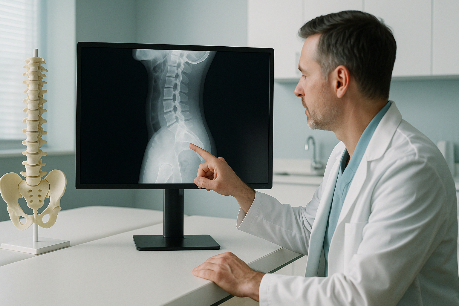
Standing, weight-bearing sagittal balance imaging captures the whole spine and pelvis in one unstitched radiograph under physiological load. This gives spinal surgeons accurate spinopelvic parameters. It shows compensation mechanisms that disappear when the patient lies down. It also supports precise planning of deformity correction and comparison after surgery.
Key takeaways
- EOS produces one unstitched full-spine standing radiograph from C2 to the femoral heads, with about 90% less radiation than a standard X-ray.
- Weight-bearing images show the true sagittal vertical axis (SVA) and compensation mechanisms such as pelvic retroversion and knee flexion.
- Spinopelvic parameters, including pelvic incidence, lumbar lordosis and PI-LL mismatch, are measured under physiological load.
- The low dose allows serial imaging for planning before surgery and comparison after surgery, without concerns about cumulative radiation.
- EOS shows bony alignment. For soft tissue, discs or nerve assessment, MRI remains the right tool.
Accurate sagittal balance assessment is central to planning adult deformity surgery. The surgical literature consistently links sagittal malalignment to poor patient-reported outcomes. Correction targets come from standing spinopelvic measurements. Yet many referral pathways still rely on supine or segmented imaging. This can understate how much imbalance the patient actually has on their feet.
At ScanAlign, unstitched full-body standing imaging at 19 Harley Street, London, we provide EOS slot-scanning radiography designed for this clinical need. The system captures the whole skeletal chain, from the skull base to below the knees, in one calibrated, weight-bearing acquisition.
Why does standing imaging change spinal surgery planning?
Standing imaging shows the spine under its true mechanical load. This is the only position that reflects the patient's functional alignment. When a patient lies supine, relaxed muscles, the absence of gravity and the support of the table can mask significant sagittal imbalance. Curves flatten, pelvic tilt reduces, and the C7 plumb line shifts posteriorly. The result is a radiograph that may underestimate the correction needed.
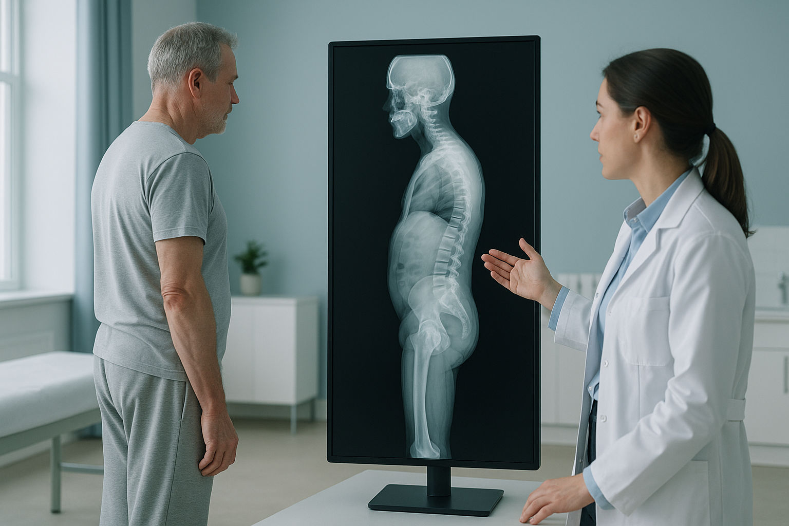
A full-spine standing radiograph with EOS removes these artefacts. The patient stands naturally and bears weight through their usual compensation strategies. If the pelvis is retroverted, the hips are flexed or the knees are bent to keep their gaze level, all of this shows in one image. This is the functional reality the surgeon must correct for.
Which spinopelvic parameters can be measured from a standing EOS scan?
All standard spinopelvic parameters used in adult deformity planning can be taken from a calibrated standing EOS acquisition. These include pelvic incidence (PI), lumbar lordosis (LL), PI-LL mismatch, sacral slope, pelvic tilt, thoracic kyphosis, the sagittal vertical axis (SVA) and the C7 plumb line relative to the sacral endplate.
The horizontal offset of C7 from the posterior sacral endplate, measured under true weight-bearing conditions.
The difference between pelvic incidence and lumbar lordosis, a core planning measure for lordosis restoration targets.
Weight-bearing pelvic tilt shows active compensation. Supine values may differ significantly from standing values.
Full-length imaging captures thoracic kyphosis, cervical lordosis and coronal alignment at the same time.
EOS captures the whole skeletal chain in one unstitched exposure. So there are no stitching artefacts that can distort angle measurements. The image is geometrically calibrated, which supports reliable output from measurement software.
How do standing and supine imaging compare for deformity assessment?
In the same patient, standing imaging consistently shows greater sagittal imbalance than supine imaging. The difference is clinically meaningful and can change surgical targets.
True physiological load. The patient stands in their natural posture, including any compensatory strategies. The SVA, pelvic tilt and knee flexion are all visible. The C7 plumb line position reflects the functional offset the patient lives with.
Unstitched, calibrated acquisition. One pass from head to below the knees captures the full chain without stitching error. The low-dose slot-scanning technology delivers about 90% less radiation than a standard X-ray, around ten times less. This makes serial imaging practical for comparison before and after surgery.
Gravity is removed. The table supports the spine, which relaxes the paraspinal muscles and reduces the curves. Pelvic tilt often decreases. The SVA shifts posteriorly, which masks how much forward imbalance there is. In patients with flatback syndrome or fixed sagittal imbalance, supine films may look misleadingly normal.
Compensation is hidden. Pelvic retroversion, hip extension and knee flexion do not show on a supine film. These compensatory mechanisms are critical to understanding how much correction is needed and where it should be placed.
In adult deformity surgery planning, the difference between standing and supine spinopelvic measurements can exceed clinical thresholds. Surgical targets taken from supine data risk under-correction. The literature links under-correction with revision surgery and poor outcomes.
How do surgeons refer patients for standing sagittal balance imaging?
Surgeons can refer patients directly to ScanAlign for EOS sagittal balance imaging. No referral from a family doctor is needed. The process is simple and designed to fit the surgical workflow.
Register as a referrer
Complete a short registration so we can link referrals to your practice. You can register to refer patients online.
Send a referral
Send us the clinical indication and any specific imaging needs. We can take requests for particular views, field of view or inclusion of the lower limbs.
The patient comes to London for imaging
The scan takes place at our clinic at 19 Harley Street, London. The acquisition itself typically lasts under a minute. The patient stands in their natural posture throughout.
Images and measurements sent to you
We provide calibrated DICOM images along with spinopelvic parameter measurements. These work with standard deformity planning software.
If you are a spinal surgeon or a member of a clinical team and would like to discuss imaging needs, you can refer a patient or contact us directly.
Book a free video consultationWhen is MRI more appropriate than EOS for spinal assessment?
MRI is usually the better tool when the clinical question involves soft tissue, discs or nerves. EOS shows bones, joints and alignment with precision under weight-bearing conditions. But it does not image the spinal cord, disc hydration, neural foramina or ligaments.
EOS and MRI work together. EOS provides the global alignment and spinopelvic parameter data needed to plan deformity correction. MRI provides the soft-tissue detail needed to plan neural decompression and assess discs. In most adult deformity cases, both modalities contribute to the surgical plan.
Before a patient starts treatment, or changes the one they are on, a detailed diagnosis is worth getting. Knowing whether the cause sits in the pelvis, the legs or the spine changes what will actually help.
Frequently asked questions about sagittal balance imaging
Sagittal balance imaging is a standing, weight-bearing radiograph that captures the whole spine and pelvis in the sagittal plane. It allows measurement of the key spinopelvic parameters used to assess sagittal malalignment and plan its correction.
The sagittal vertical axis reflects the patient's functional balance under gravity. When the patient lies down, gravity is removed, muscles relax, and the SVA shifts posteriorly. Measuring in the standing position captures the true offset and any compensation mechanisms the patient uses.
EOS uses slot-scanning technology to produce one unstitched, calibrated image from head to feet. Conventional full-length standing X-rays are typically stitched together from several cassettes, which can introduce measurement error. EOS also delivers about 90% less radiation than a standard X-ray, around ten times less.
Yes. The calibrated EOS image includes the pelvis and the full lumbar spine. This allows accurate measurement of both pelvic incidence and lumbar lordosis. The PI-LL mismatch is calculated from these values.
EOS delivers about 90% less radiation than a standard X-ray, around ten times less. This low dose makes it practical for serial imaging for planning before surgery and comparison after surgery, without the cumulative radiation concerns linked with conventional radiography or CT.
No. Consultant surgeons can refer patients directly. Patients may also refer themselves. There is no need for a family doctor to be involved.
Common indications include adult spinal deformity assessment, flatback syndrome, degenerative sagittal imbalance, planning before corrective osteotomy or long fusion, monitoring of alignment after surgery, and assessment of adjacent segment disease after previous surgery.
Yes. EOS images are delivered as calibrated DICOM files. They work with standard surgical planning software used for adult deformity correction, including tools that calculate correction targets from spinopelvic parameters.
The acquisition itself typically lasts under a minute. The patient stands still in their natural posture while the scanner moves from head to feet. The whole appointment, including positioning, is brief.
ScanAlign is a private, self-pay clinic. The price for patients travelling from abroad is confirmed during the free video consultation.
Refer a patient for standing sagittal balance imaging
If you would like to discuss a case or refer a patient for EOS imaging at our clinic at 19 Harley Street, London, we are happy to help.
Book a free video consultationWritten by Abbas Dhami (Specialist Diagnostic Radiographer)
Sources
- Schwab F, Ungar B, Blondel B, et al. Scoliosis Research Society-Schwab adult spinal deformity classification: a validation study. Spine. 2012;37(12):1077-1082.
- Lazennec JY, Brusson A, Rousseau MA. Lumbar-pelvic-femoral balance on sitting and standing lateral radiographs. Orthopaedics & Traumatology: Surgery & Research. 2013;99(1 Suppl):S87-S103.
- Deschênes S, Charron G, Beaudoin G, et al. Diagnostic imaging of spinal deformities: reducing patients radiation dose with a new slot-scanning X-ray imager. Spine. 2010;35(9):989-994.
This page is general information and not a diagnosis.
ScanAlign is the trading name of CoreMed Solutions Ltd and operates under The Harley Street Hospital's CQC licence.