

What Is Sacroiliitis, and How Is It Different From SI Joint Dysfunction?
Sacroiliitis is inflammation inside the sacroiliac (SI) joint. It is often linked to an autoimmune or inflammatory condition. SI joint dysfunction is a mechanical problem, where the joint moves too much or too little. Both cause pain in the buttock and lower back. But they have different causes, need different tests and respond to different approaches.
Key points
- Sacroiliitis means the SI joint is inflamed, not just stiff or out of line.
- It is commonly linked with axial spondyloarthritis and other inflammatory conditions.
- MRI is the best imaging test for finding active inflammation (bone marrow oedema) in the SI joint.
- Standing alignment imaging can help rule out mechanical causes, such as a difference in leg length or a pelvic tilt.
- If your symptoms include night pain, morning stiffness lasting over 30 minutes, or buttock pain that switches sides, see your doctor for blood tests and a possible referral to a rheumatologist.
The sacroiliac joints sit at the back of your pelvis. They connect your spine to your hip bones. They carry a huge amount of load every time you stand, walk or climb stairs. When something goes wrong here, the pain can be confusing. It often feels like lower back pain, deep pain in the buttock, or even pain down the leg.
The hard part is working out whether the pain comes from inflammation or from a mechanical problem. This matters, because the answer changes everything about how the problem is managed.
What are the symptoms of sacroiliitis?
Sacroiliitis usually causes a deep, aching pain in the lower back or buttocks. It is worse after rest and gets better with movement. The classic pattern is morning stiffness that lasts more than 30 minutes. It sometimes wakes you at night.
Other symptoms can include tiredness, a general feeling of being unwell and, at times, pain spreading down the back of the thigh. Because the pain is in the same area, it is sometimes mistaken for ischial bursitis or sciatica.
Get urgent medical help if you develop numbness or weakness in both legs, or any loss of bladder or bowel control. Contact your doctor or go to the nearest emergency department straight away.
What causes sacroiliitis?
Sacroiliitis is caused by inflammation inside the SI joint itself. The most common cause is an autoimmune or inflammatory condition. Infection and mechanical overload can also trigger it.
A blood marker called HLA-B27 is found in many people with axial spondyloarthritis. But not everyone who carries it develops the condition. Your doctor or rheumatologist can arrange this blood test, along with inflammatory markers such as CRP and ESR.
How is sacroiliitis different from SI joint dysfunction?
The main difference is the cause of the pain. Sacroiliitis is driven by inflammation inside the joint. SI joint dysfunction is a mechanical problem. The joint is stiff, hypermobile, or sitting out of its normal position because of a pelvic tilt, a difference in leg length or a muscle imbalance.
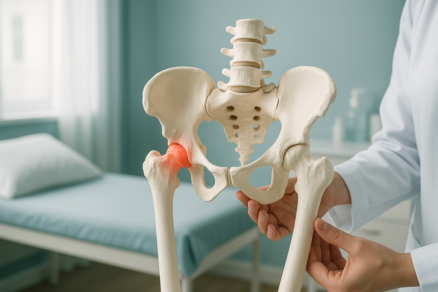
In real life, the two can overlap. A pelvis that sits unevenly puts extra stress on one SI joint, which may then become inflamed. That is why looking at both alignment and inflammation gives a clearer answer than either one alone.
How is sacroiliitis diagnosed with imaging?
MRI is the gold-standard imaging test for finding active sacroiliitis. It can show bone marrow oedema, the telltale sign of active inflammation inside the SI joint. It shows this long before structural damage appears on a standard X-ray.
When MRI is the better test. To confirm active sacroiliitis, MRI is usually essential. It shows soft tissue, bone marrow oedema and inflammation that X-ray and CT cannot detect. If your clinician suspects an inflammatory cause, an MRI of the SI joints should come first.
Where standing imaging fits in
Your pain pattern may sound more mechanical, or MRI may have already ruled out active inflammation. Then the next question is whether your pelvis and spine are aligned under load. A standard X-ray or MRI is taken lying down. Lying flat relaxes your muscles and removes the effect of gravity. So it can hide alignment problems that only show up when you are standing and bearing weight.
ScanAlign standing EOS imaging captures your full skeleton from head to toe while you stand naturally. It uses about 90% less radiation than a standard X-ray, around ten times less. This kind of image can show whether a pelvic tilt, a difference in leg length or a spinal curve is overloading one SI joint and adding to your pain.
Not sure whether your SI joint pain is inflammatory or mechanical? A free video consultation, from your home, can help you understand which type of imaging would be most useful before you plan a trip to London.
Book a free video consultationWhat helps manage sacroiliitis?
Management depends entirely on the cause. ScanAlign is a diagnostic imaging clinic and does not provide treatment. But knowing the broad approaches can help you have a better-informed talk with your doctor or specialist.
- Referral to a rheumatologist for diagnosis and decisions on medicine
- Anti-inflammatory medicine or targeted biological therapies
- Structured exercise programmes to keep you mobile
- Ongoing monitoring with MRI and blood tests
- Physiotherapy aimed at muscle imbalances and pelvic stability
- Assessment of alignment, leg-length difference or pelvic tilt
- Changes in footwear or orthotics if a difference in leg length is confirmed
- Strengthening the muscles around the pelvis and core
The right starting point is the right diagnosis. If your symptoms sound inflammatory, ask your doctor about HLA-B27 testing, inflammatory blood markers and a referral to a rheumatologist. If they sound mechanical, standing alignment imaging can show what lying-down scans miss. You can book a standing pelvis scan to look at your alignment under load.
Before you start a treatment, or change the one you are on, it is worth getting a detailed diagnosis. Knowing whether the cause sits in your pelvis, your legs or your spine changes what will actually help.
Frequently asked questions about sacroiliitis
Not exactly. Sacroiliitis is inflammation of the SI joint. Ankylosing spondylitis is one specific inflammatory condition that commonly causes sacroiliitis. Other conditions, such as psoriatic arthritis and reactive arthritis, can cause it too.
Yes. Early inflammatory sacroiliitis sometimes affects one side first before spreading to both. Mechanical causes can also cause one-sided symptoms, especially if there is a pelvic tilt or a difference in leg length.
Alternating buttock pain is pain that switches sides over time. It is seen as a classic feature of inflammatory sacroiliitis and axial spondyloarthritis. If you notice this pattern, mention it to your doctor.
Standard X-rays can show long-term changes such as erosions or fusion, but they often miss early active inflammation. MRI is far more sensitive for finding bone marrow oedema, the hallmark of active sacroiliitis. It is the recommended first-line imaging test.
Bone marrow oedema is fluid and swelling inside the bone near the joint surface. On MRI, it shows up as a bright signal. It is the key sign that active inflammation is present in the SI joint.
No. ScanAlign accepts self-referrals. You can book a free video consultation directly to discuss whether standing alignment imaging is right for your situation.
An EOS scan uses about 90% less radiation than a standard X-ray, around ten times less. This makes it suitable for full-body standing images without significant radiation exposure.
The price for patients travelling from abroad is confirmed during the free video consultation. This consultation before booking also helps make sure you get the right scan for your needs.
It can. Pain from the SI joint sometimes spreads down the back of the thigh, and at times to the calf. This can be confused with sciatica, which is why an accurate diagnosis matters.
If your symptoms are inflammatory, with morning stiffness, night pain and improvement with movement, a rheumatologist is usually the right specialist. If symptoms are more mechanical, with pain linked to posture, load and specific movements, an orthopaedic opinion or physiotherapy may be more suitable.
Unsure what is causing your SI joint pain?
A free video consultation can help you understand whether standing alignment imaging, MRI or both would give you the clearest answers.
Book a free video consultationWritten by Abbas Dhami (Specialist Diagnostic Radiographer)
Sources
- Sieper J, Poddubnyy D. Axial spondyloarthritis. The Lancet. 2017;390(10089):73-84.
- National Institute for Health and Care Excellence (NICE). Spondyloarthritis in over 16s: diagnosis and management (NG65). 2017.
- Rudwaleit M et al. The Assessment of SpondyloArthritis International Society (ASAS) classification criteria for axial spondyloarthritis. Annals of the Rheumatic Diseases. 2009;68(6):777-783.
This page is general information and not a diagnosis.
ScanAlign is the trading name of CoreMed Solutions Ltd and operates under The Harley Street Hospital's CQC licence.