

Ischial Bursitis: What Causes Sit-Bone Pain and How Can You Find Relief?
Ischial bursitis is inflammation of the small fluid-filled sac (bursa) over your ischial tuberosity, the bony point you sit on. It causes a deep, aching pain in the buttock. The pain gets worse when you sit on hard surfaces. The most common triggers are long periods of pressure, repetitive strain, or problems with pelvic alignment.
Key points
- Ischial bursitis, sometimes called weaver's bottom or tailor's bottom, is inflammation of the bursa over your sit bone.
- The pain is usually worst on hard chairs. It may spread down the back of the thigh.
- Pelvic tilt and differences in leg length can overload one sit bone, so symptoms keep going.
- A standing, weight-bearing scan can show alignment factors that lying-down imaging misses.
- EOS imaging uses about 90% less radiation than a standard X-ray, around ten times less.
You may have heard it called ischiogluteal bursitis, weaver's bottom or tailor's bottom. These names come from trades where people sat for long hours on hard benches. The problem is the same today. It affects anyone who spends long periods sitting, cycling or running.
Normally the bursa acts as a cushion between the bone and the tendons attached around it. When it becomes irritated, you press straight onto the inflamed tissue every time you sit down. That is why the pain can feel so sharp and so exact in one spot.
What does ischial bursitis feel like?
The main sign is a deep ache in one clear spot, right on the sit bone. It often gets worse when you sit for a long time and eases when you stand or walk. Some people also feel pain when climbing stairs or stretching the hamstrings.
A sharp or burning ache centred on one buttock, right over the bony point.
Sitting on a wooden chair, a bench or a bicycle saddle makes the discomfort clearly worse.
The ache can spread along the back of the upper leg. It can feel like a hamstring problem.
Getting up after sitting for a while feels stiff. The pain eases once you move around.
Ischial bursitis and hamstring tendinopathy affect almost the same area, so people often mix them up. Ischial tuberosity bursitis tends to hurt most with direct pressure on the bone. Hamstring tendinopathy is usually worse during active movement, such as running or lunging. A thorough assessment helps tell them apart.
What causes ischial bursitis?
Ischial bursitis develops when repeated friction or pressure irritates the bursa. Sitting for long periods on hard surfaces is the most common trigger. But alignment and activity patterns play a bigger role than many people realise.
Desk work, long drives or sitting on hard benches press the bursa against the bone for hours.
Cycling, rowing or running can load the ischial area again and again and inflame the bursa.
An uneven pelvis shifts more weight onto one sit bone. This overloads the bursa on that side.
Tight hamstrings or gluteal muscles increase friction across the bursa with every movement.
How does alignment affect sit-bone pain?
Your pelvis is the base of your sitting posture. If it tilts or rotates even slightly, one ischial tuberosity carries more load than the other. Over weeks and months, that imbalance can keep the bursa inflamed, however many cushions you try.
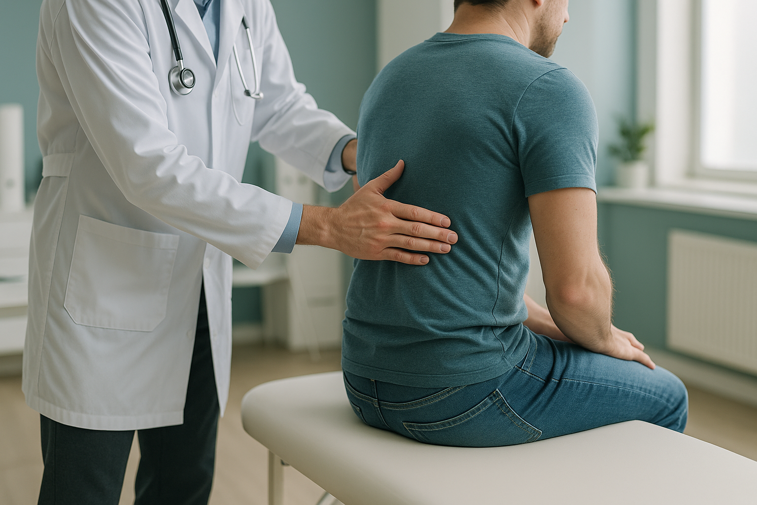
The body works as one connected chain, from the feet to the head. A difference in leg length, a rotated hip or a curve in the lower spine can all shift the pelvis out of balance. The pain shows up at the sit bone, but the root cause may be somewhere else entirely.
This is why standard imaging of the pelvis alone does not always give the full picture. Seeing the whole chain, from your feet up through your spine, shows compensations that a scan of one area would miss.
How can a standing EOS scan help with ischial bursitis?
A standing EOS scan captures your whole skeleton while you stand upright and bear your own weight. So it shows your pelvis, hips, legs and spine as they work when you sit and stand, not relaxed on a table.
Lying-down imaging, such as a standard X-ray or CT, relaxes the muscles. It lets the pelvis settle into a neutral position. Alignment problems that only appear under gravity can be invisible on those scans. A standing image captures what really happens when your body carries its own weight.
EOS uses about 90% less radiation than a standard X-ray, around ten times less. It produces a full-length image in a single pass. Your clinician can then measure pelvic tilt, leg-length differences and spinal alignment, all from one scan. That information helps explain why one side may be taking more pressure than the other.
Wondering if your sit-bone pain could be linked to alignment? A short video call can help you decide if imaging is the right next step, before you plan a trip to London.
Book a free video consultationWhen MRI is the better choice: EOS shows bones, joints and alignment clearly. If your clinician suspects a soft-tissue problem, such as hamstring tendon damage, a labral tear or nerve irritation, MRI is usually the better tool for seeing those structures in detail.
What helps manage ischial bursitis?
Most people find relief by reducing direct pressure on the area and dealing with any underlying causes. Your doctor or physiotherapist can guide you on the best approach for you. Common steps include:
- Cushioned seating. A pressure-relieving cushion takes load off the ischial tuberosity while you sit.
- Changing your activities. Spending less time on hard surfaces and adjusting your cycling or rowing technique can calm the inflammation.
- Stretching and strengthening. Gentle hamstring and gluteal exercises, guided by a physiotherapist, can reduce tension across the bursa.
- Alignment assessment. If a pelvic tilt or a difference in leg length is playing a part, finding it and addressing it may stop the problem coming back.
ScanAlign is a diagnostic imaging clinic. It does not provide treatment. But the detailed alignment data from a standing scan can give your treating clinician the information they need to target the cause, not just the symptoms.
Before you start a treatment, or change the one you are on, it is worth getting a detailed diagnosis. Knowing whether the cause sits in your pelvis, your legs or your spine changes what will actually help.
You can learn more about how standing whole-body imaging works on the EOS scan overview page.
Frequently asked questions about ischial bursitis
Ischial bursitis is inflammation of the bursa, a small fluid-filled cushion, that sits over the ischial tuberosity (your sit bone). It causes a deep ache in the buttock that is often worst when sitting on hard surfaces.
Yes. Weaver's bottom, tailor's bottom and ischiogluteal bursitis are all names for the same condition. The names come from jobs that involved long periods of sitting on hard surfaces.
Both cause pain at the sit bone. Ischial bursitis tends to hurt most with direct pressure, such as sitting. Hamstring tendinopathy is usually worse during active movements like running, lunging or stretching. A clinical assessment can help tell them apart.
Yes. If the pelvis tilts or rotates, one sit bone carries more weight than the other. That uneven load can irritate the bursa on the heavier side and keep symptoms going even after rest.
An EOS scan takes a full-length image of your skeleton while you are standing. This shows your pelvis, legs and spine while they carry your real weight. A standard X-ray usually captures only one area and is often taken lying down, which can hide alignment problems.
EOS uses about 90% less radiation than a standard X-ray, around ten times less. You can find more details on safety on the FAQs page.
No. ScanAlign accepts self-referrals. You can book a free video consultation directly to discuss whether imaging is right for your situation.
The price for patients travelling from abroad is confirmed during the free video consultation. You can find more on the FAQs page.
ScanAlign provides diagnostic imaging, not treatment. The scan report gives detailed alignment data that your treating clinician, whether a physiotherapist, osteopath or orthopaedic surgeon, can use to guide your care.
If you notice numbness, weakness in the leg, or any loss of bladder or bowel control along with your pain, seek urgent medical attention from your doctor or the nearest emergency department. These symptoms need prompt assessment.
Still unsure what is causing your sit-bone pain?
A free video consultation lets you talk through your symptoms from home and find out whether a standing alignment scan in London could help.
Book a free video consultationWritten by Abbas Dhami (Specialist Diagnostic Radiographer)
Sources
- NHS, "Bursitis," nhs.uk, accessed 2024.
- Wisniewski SJ, Grogg B. "Ischial bursitis," UpToDate, Wolters Kluwer, accessed 2024.
- Dubousset J et al. "EOS stereo-radiography system: whole-body simultaneous anteroposterior and lateral radiographs with very low radiation dose," Revue de Chirurgie Orthopédique et Réparatrice de l'Appareil Moteur, 2005; 91(5 Suppl): 13-25.
This page is general information and not a diagnosis.
ScanAlign is the trading name of CoreMed Solutions Ltd and operates under The Harley Street Hospital's CQC licence.