

What Is the Risser Sign, and How Is Skeletal Maturity Measured?
The Risser sign is a grading system. It estimates how much spinal growth a young person has left. It looks at the bony cap (apophysis) on the top of the pelvis on an X-ray. It is graded from 0 to 5, and a higher number means less growth is left. Clinicians use it to guide monitoring and treatment decisions for scoliosis.
Key takeaways
- The Risser sign grades skeletal maturity from 0 (still growing) to 5 (growth complete).
- It is measured on a standard pelvis X-ray, by looking at the iliac crest apophysis.
- Low Risser grades carry a higher risk of the scoliosis curve getting worse.
- Sanders staging of the hand and wrist gives a more detailed view of how much growth is left.
- Standing, weight-bearing imaging shows the pelvis and spine under real load.
How does the Risser sign work?
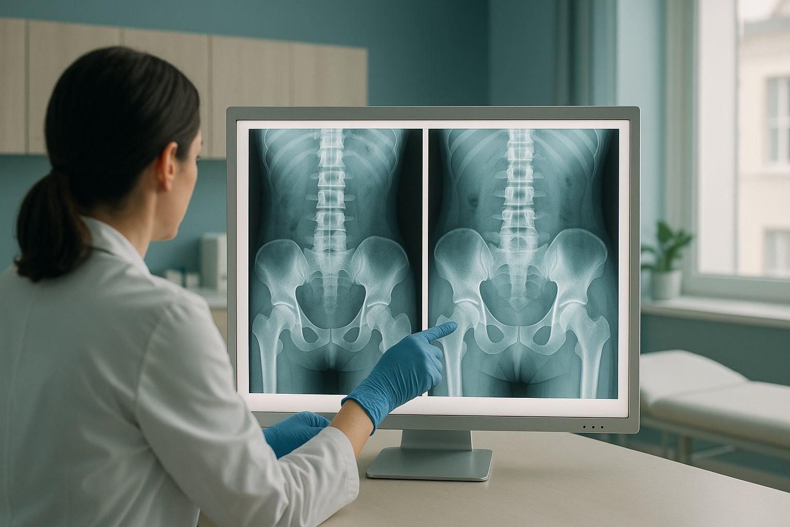
The Risser sign tracks a strip of cartilage along the top rim of the pelvis. This strip is called the iliac apophysis. During the teenage years, this cartilage slowly turns into bone. The change moves from the outer hip towards the spine. A radiographer or radiologist looks at this strip on an X-ray. They give a grade from 0 to 5, based on how far the ossification has gone and whether it has fused to the pelvis.
The pelvis almost always shows on a full-spine X-ray. So the Risser sign can be checked at the same time as a Cobb angle measurement, with no need for a separate scan.
What do the Risser grades mean?
Each Risser grade describes a set stage of bone maturity in the pelvis. The higher the grade, the less growth is left.
No ossification can be seen. The skeleton is still in an early growth phase. The risk of the curve getting worse is highest at this stage.
Ossification covers the outer quarter of the iliac crest. A lot of growth is still left.
Ossification reaches the halfway point. Growth is slowing, but the skeleton is not yet mature.
Ossification covers three quarters of the crest. Most of the fast growth has passed, but some is left.
The apophysis runs the full length of the crest but has not yet fused to the pelvis. Very little growth is left.
Complete fusion. Skeletal growth is considered finished. The risk of the curve getting worse is much lower.
What is Sanders staging and how does it compare to the Risser sign?
Sanders staging is a newer system. It uses a hand and wrist X-ray to check bone age in more detail. It splits skeletal maturity into eight stages. This gives clinicians finer detail during the fast growth phase, when the risk of the curve getting worse is greatest.
The Risser sign is useful but fairly broad. A patient can stay at Risser 0 for a long time, and a scoliosis curve may change a lot during this time. Sanders staging helps show exactly where the patient is in the growth spurt. This makes it easier to decide whether bracing, closer monitoring or watchful waiting is right.
In practice, many specialists use both systems together. A spine X-ray gives the Risser grade and the Cobb angle measurement. A separate hand X-ray gives the Sanders stage.
What it looks at: The bony cap on the pelvis (iliac crest apophysis).
Scale: 0 to 5.
Strength: Easy to read on a standard spine or pelvis X-ray. Widely understood by clinicians.
Limitation: Less precise during the fastest growth period (Risser 0 to 1).
What it looks at: The growth plates in the hand and wrist bones.
Scale: 1 to 8.
Strength: Finer detail during peak growth, which helps predict how the curve will behave more precisely.
Limitation: Needs a separate hand X-ray. Less widely used outside specialist scoliosis centres.
Why does skeletal maturity matter for scoliosis?
Skeletal maturity matters because a spine that is still growing is at far greater risk of a scoliosis curve getting worse. The more growth is left, the more time the curve has to progress. That is why clinicians check skeletal maturity at every monitoring appointment.
A low Risser grade together with a larger curve is a strong sign that treatment, usually bracing, may be needed to slow or prevent the curve getting worse. A high Risser grade with a stable curve often means the patient can be checked less often, or discharged from active monitoring altogether.
- Risser 0 or 1
- Larger curve size
- Before or during the growth spurt
- Sanders stage 1 to 3
- Risser 4 or 5
- Smaller, stable curve
- Growth spurt already passed
- Sanders stage 7 or 8
When is MRI a better choice? The Risser sign and Cobb angle are measured on X-ray, which shows bones, joints and alignment clearly. If there is concern about the spinal cord, nerves or discs, for example with unusual pain, fast progression or neurological symptoms, MRI is usually the better tool. This is because it shows soft tissue in detail.
How does standing imaging help with skeletal maturity assessment?
Standing, weight-bearing imaging shows the spine and pelvis exactly as they behave when a person is upright. When you lie down for a normal scan, gravity is taken away and muscles relax. Curves can then look smaller than they really are. A standing X-ray shows the real load on the skeleton. It gives the most accurate view of both the curve and the Risser sign in one image.
At ScanAlign, low dose standing imaging, full-length standing scans use about 90% less radiation than a standard X-ray, around ten times less. Scoliosis needs repeat checks over months or years, so keeping the radiation dose as low as possible matters.
Before you start treatment, or change the treatment you are on, it is worth getting a detailed diagnosis. Knowing whether the cause sits in the pelvis, the legs or the spine changes what will actually help.
Do you have a scoliosis monitoring report that mentions the Risser sign? If you want to understand what it means for your next steps, a free video consultation from home can help you decide whether a standing scan in London is right for you.
Book a free video consultationFrequently asked questions about the Risser sign
Risser 0 means no ossification can be seen along the iliac crest. The skeleton is still in an early growth phase. This means there is a higher risk that a scoliosis curve could get worse. Closer monitoring is usually advised at this stage.
At Risser 3, the fastest growth has usually passed, but some growth is left. Curves may still change, so monitoring usually continues until the Risser grade reaches 4 or 5.
Scoliosis monitoring is the most common use. But orthopaedic surgeons also look at skeletal maturity for other growth-related conditions, and when planning the timing of surgery.
MRI shows soft tissue best. The Risser sign is checked on a plain X-ray, because it depends on seeing bone ossification clearly. An MRI of the pelvis may show the apophysis, but X-ray is still the standard method.
The Risser sign looks at the pelvis and uses a 0 to 5 scale. Sanders staging looks at the hand and wrist and uses a 1 to 8 scale. Sanders gives more detail during fast growth phases. The Risser sign is more widely used and can be read on any standard spine X-ray.
Your specialist will set the monitoring schedule based on your curve size and growth stage. It is usually every four to six months during peak growth, though this varies from person to person.
The Risser grade itself does not change between standing and lying down, because it measures bone maturity, not alignment. But a standing X-ray gives a more accurate picture of the scoliosis curve at the same time. This matters when the Risser grade and the curve are assessed together.
No. ScanAlign offers self-referred appointments for adults. You can book a free video consultation to discuss whether a standing scan is right for your situation.
The EOS system used at ScanAlign delivers about 90% less radiation than a standard X-ray, around ten times less. This is especially important when repeat scans are needed over time.
The price for patients travelling from abroad is confirmed during the free video consultation. This consultation, before booking, also helps confirm the right scan for your needs.
Understand your Risser grade in context
A free video consultation with our team can help you understand your report, judge whether a standing scan would add useful information, and plan your next steps before you travel.
Book a free video consultationSources
- Risser JC. The iliac apophysis: an invaluable sign in the management of scoliosis. Clinical Orthopaedics and Related Research. 1958;11:111-119.
- Sanders JO et al. Maturity assessment and curve progression in girls with idiopathic scoliosis. Journal of Bone and Joint Surgery (American). 2007;89(1):64-73.
- Scoliosis Research Society. Patient information: skeletal maturity and scoliosis. srs.org.
Written by Abbas Dhami (Specialist Diagnostic Radiographer)
This page is general information and not a diagnosis.
ScanAlign is the trading name of CoreMed Solutions Ltd and operates under The Harley Street Hospital's CQC licence.