
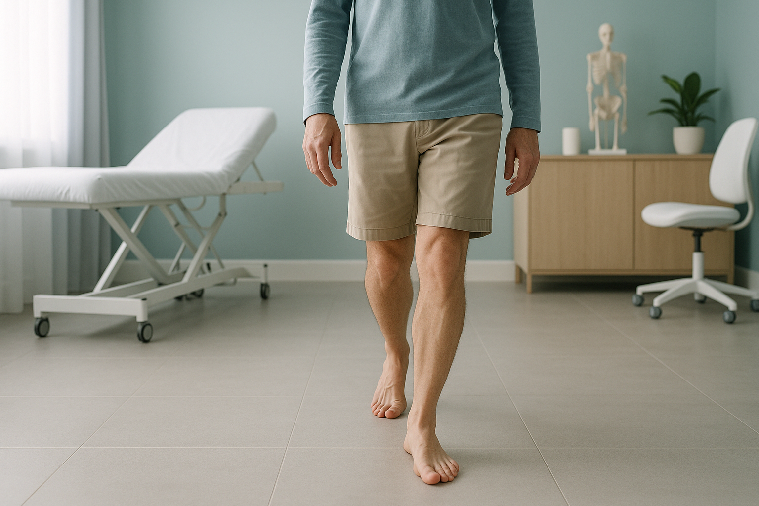
Why Do I Walk With My Feet Turned Out?
Out-toeing in adults, sometimes called duck feet walking, is when one or both feet point outward instead of straight ahead. It is usually caused by the shape of your hip or shin bones, or by tightness and weakness around the hip. A whole-body standing scan can show the exact rotational profile of your legs, so the right approach can be chosen.
Key points
- Out-toeing is a rotation pattern, not just a foot problem. The cause often sits at the hip or shin.
- Common structural causes include femoral retroversion and external tibial torsion.
- Lying-down scans relax the legs. This hides how rotation really loads the joints when you stand and walk.
- A standing EOS scan maps the full rotational profile from hip to ankle in one low-dose image.
- Knowing the cause helps clinicians decide whether physiotherapy, orthotics or further tests are needed.
What is out-toeing in adults?
Out-toeing is the medical term for a walking pattern where one or both feet point outward, rather than facing forwards. You may know it as "duck feet". In children it often goes away on its own. In adults it tends to be a fixed pattern, linked to your bone structure, how far your joints can move, or long-standing muscle imbalances.
The direction your foot points as you walk is called the foot progression angle. When that angle points clearly outward, the rotation has to come from somewhere higher up the leg. It might start at the hip socket, the thigh bone, the shin bone or the foot itself. Often it comes from more than one level.
How do you know if you have out-toeing?
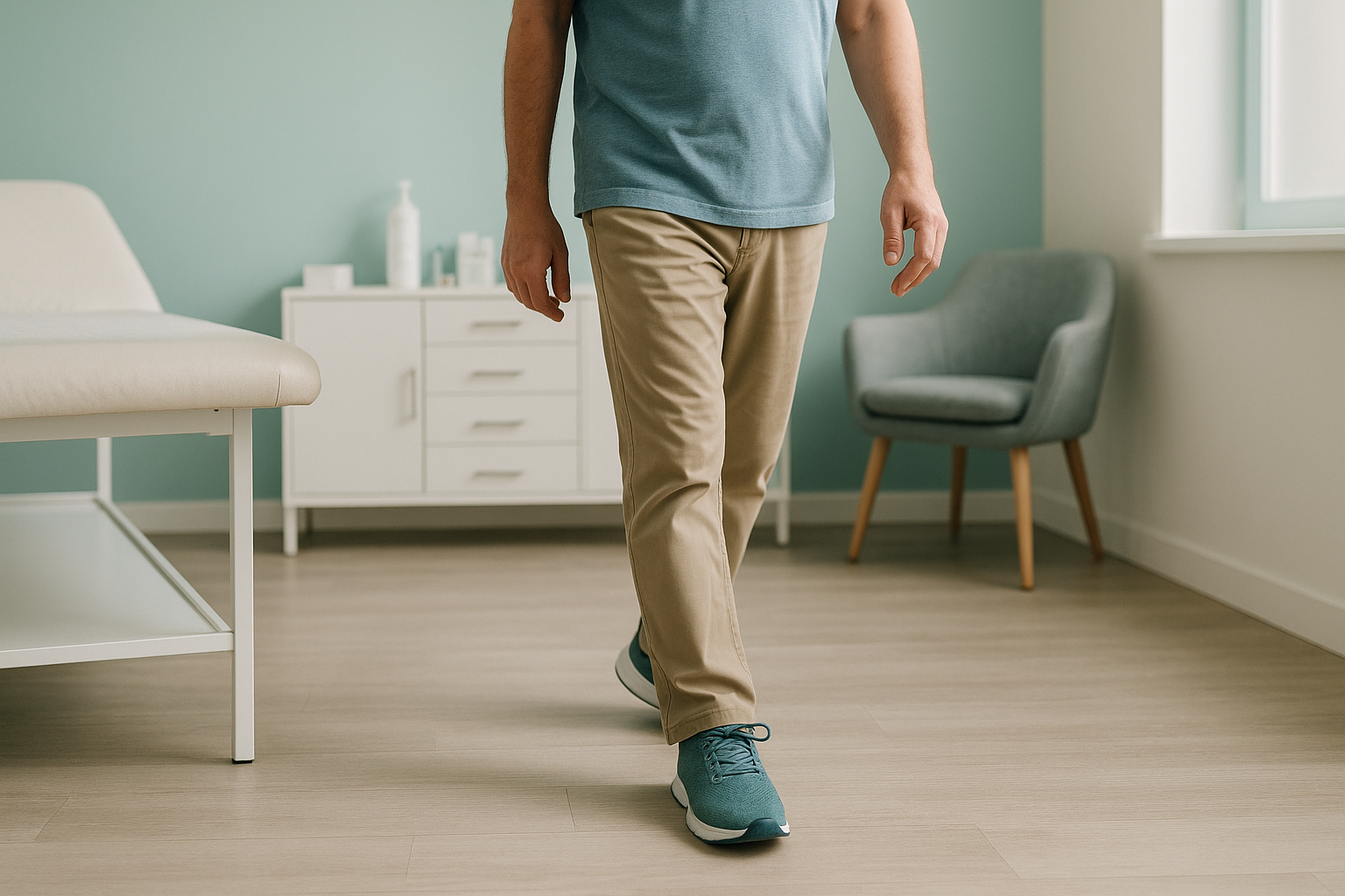
You may have noticed it yourself, or someone else may have pointed it out. These are the most common signs.
When you stand or walk, your feet clearly point outward rather than straight ahead.
The inner edges of your soles wear down faster because of the turned foot position.
Pain or tiredness in the hip, knee or ankle after walking or standing for a while, caused by compensatory loading.
Your legs flick outward when you run. This makes running less efficient and sometimes causes tripping.
What causes out-toeing in adults?
Out-toeing is usually structural. This means the bones themselves are shaped in a way that turns the leg outward. Sometimes tight soft tissue or joint changes play a part too. These are the most common causes.
The thigh bone is twisted, so the hip naturally sits more turned out. This is one of the most common structural causes. Learn more about anteversion versus retroversion.
The shin bone is turned outward compared with the thigh bone. This points the foot outward even when the hip is in a neutral position.
Tight external rotator muscles or a stiff hip capsule can limit internal rotation, forcing the leg outward. Read about hip anteversion and retroversion.
Collapsed arches can make the foot roll inward and splay outward. This adds to the out-toed look.
In many cases, more than one factor is present. For example, mild femoral retroversion together with external tibial torsion can produce a clearly turned-out walk, even though neither factor alone would stand out. This is why a full rotational profile, measured from hip to ankle, gives a much clearer picture than looking at one joint on its own.
How is out-toeing assessed with standing imaging?
A standing, weight-bearing scan captures your skeleton the way it really works: under gravity, with your muscles working. When you lie down for a normal X-ray, CT or MRI, your muscles relax and your joints settle into a different position. Rotation patterns that are obvious when you walk can almost disappear on a lying-down image.
An EOS scan at ScanAlign, 3D standing skeletal imaging, captures both full legs in one upright image. Because you are standing naturally, the image shows the true rotational alignment of each bone, from the hip socket down to the ankle. A 3D reconstruction can then map the exact twist of the femur and tibia. This gives clinicians a complete rotational profile to guide decisions.
The scan uses about 90% less radiation than a standard X-ray, around ten times less.
Standard X-rays, CT and MRI are taken lying down. Without gravity and working muscles, rotation at the hip, knee and ankle can be hidden. CT can measure bone twist using special slices, but it gives a higher radiation dose. It still does not show how the body takes load in a standing position.
Your body is a connected chain. If the femur sits more turned out than usual, the knee, shin and foot all have to compensate. Pain often shows up away from the root cause. A gait analysis together with a standing rotational scan gives both the movement picture and the structural explanation behind it.
When is MRI better? EOS is excellent at showing bone alignment, joint spacing and rotational geometry. If your clinician suspects soft-tissue damage, a labral tear, cartilage injury or nerve involvement, MRI is usually the better tool. The two tests work well together.
What helps with out-toeing once you know the cause?
The right approach depends entirely on what is driving the rotation. That is why accurate imaging matters before any plan is made. ScanAlign is a diagnostic imaging clinic, so it does not provide treatment. But the information from a standing scan helps your clinician choose the best path.
- Physiotherapy and strengthening. If the out-toeing is partly driven by tight or weak muscles, a targeted programme can improve how far the hip turns inward and how you walk.
- Orthotics or footwear changes. When flat feet play a part, custom insoles may help control the position of the foot and reduce compensatory rotation higher up the leg.
- Monitoring. If imaging shows a mild structural variation with no pain or limits on what you can do, the best course may simply be reassurance and regular review.
- Referral to a surgeon. In rare cases where significant femoral retroversion or tibial torsion causes persistent pain or instability, an orthopaedic surgeon may discuss corrective options. Detailed rotational measurements from a standing scan are essential for planning.
Before you start a treatment, or change the one you are on, it is worth getting a detailed diagnosis first. Knowing whether the cause sits in the pelvis, the legs or the spine changes what will actually help.
Want to know exactly where the rotation is coming from? A standing rotational scan maps the full picture from hip to ankle.
Book a free video consultationFrequently asked questions about out-toeing in adults
Yes. "Duck feet" is a common everyday term for out-toeing, where one or both feet point outward when standing and walking. The medical term describes the same pattern.
Femoral retroversion is a structural twist of the thigh bone that sets the hip more turned out than average. It is one of the most common bone-related causes of out-toeing in adults.
It can. When the leg turns outward, the knee and hip joints take load differently. Over time this changed loading may contribute to pain, cartilage wear or compensatory muscle tiredness in those areas.
If the cause is mainly tight or weak muscles, targeted physiotherapy can improve symptoms and the way you walk. If the cause is in the bone, such as femoral retroversion or tibial torsion, exercises cannot change bone shape but may help manage symptoms.
Rotation is best seen under load. When you lie down, your muscles relax and your joints settle, which can hide the rotation pattern. A standing scan shows how your bones are really aligned when gravity, muscle activity and body weight are all at work.
Yes. EOS uses about 90% less radiation than a standard X-ray, around ten times less. It is a very low-dose imaging method. You can find more detail on our book a rotation and alignment scan page.
No. ScanAlign accepts self-referrals. You can book directly without needing a letter from your doctor.
They can contribute. Collapsed arches may make the foot roll inward and splay outward, so the out-toed look becomes more obvious. A full-leg standing image helps separate the part coming from the foot from rotation higher up the leg.
Out-toeing that is uneven is worth looking into, because it suggests a difference between the two sides in bone shape, hip movement or muscle balance. One-sided patterns often cause compensatory changes in the pelvis, spine or the other leg.
The price for patients travelling from abroad is confirmed during the free video consultation. You can find more on the book a rotation and alignment scan page.
Find out what is causing your out-toeing
A free video consultation lets you talk through your concerns with a specialist radiographer from wherever you are, and find out whether a standing rotational scan in London is the right next step.
Book a free video consultationWritten by Abbas Dhami (Specialist Diagnostic Radiographer)
Sources
- Staheli, L.T. "Rotational problems in children." Journal of Bone and Joint Surgery (American Volume), 1993.
- Guenoun, B. et al. "Reliability of a new method for lower-extremity measurements based on stereoradiographic three-dimensional reconstruction." Orthopaedics & Traumatology: Surgery & Research, 2012.
- Royal College of Radiologists. iRefer Guidelines: Making the Best Use of Clinical Radiology. RCR, London.
This page is general information and not a diagnosis.
ScanAlign is the trading name of CoreMed Solutions Ltd and operates under The Harley Street Hospital's CQC (Care Quality Commission) licence.