
Knee Anatomy: What Every Ligament and Bone Actually Does
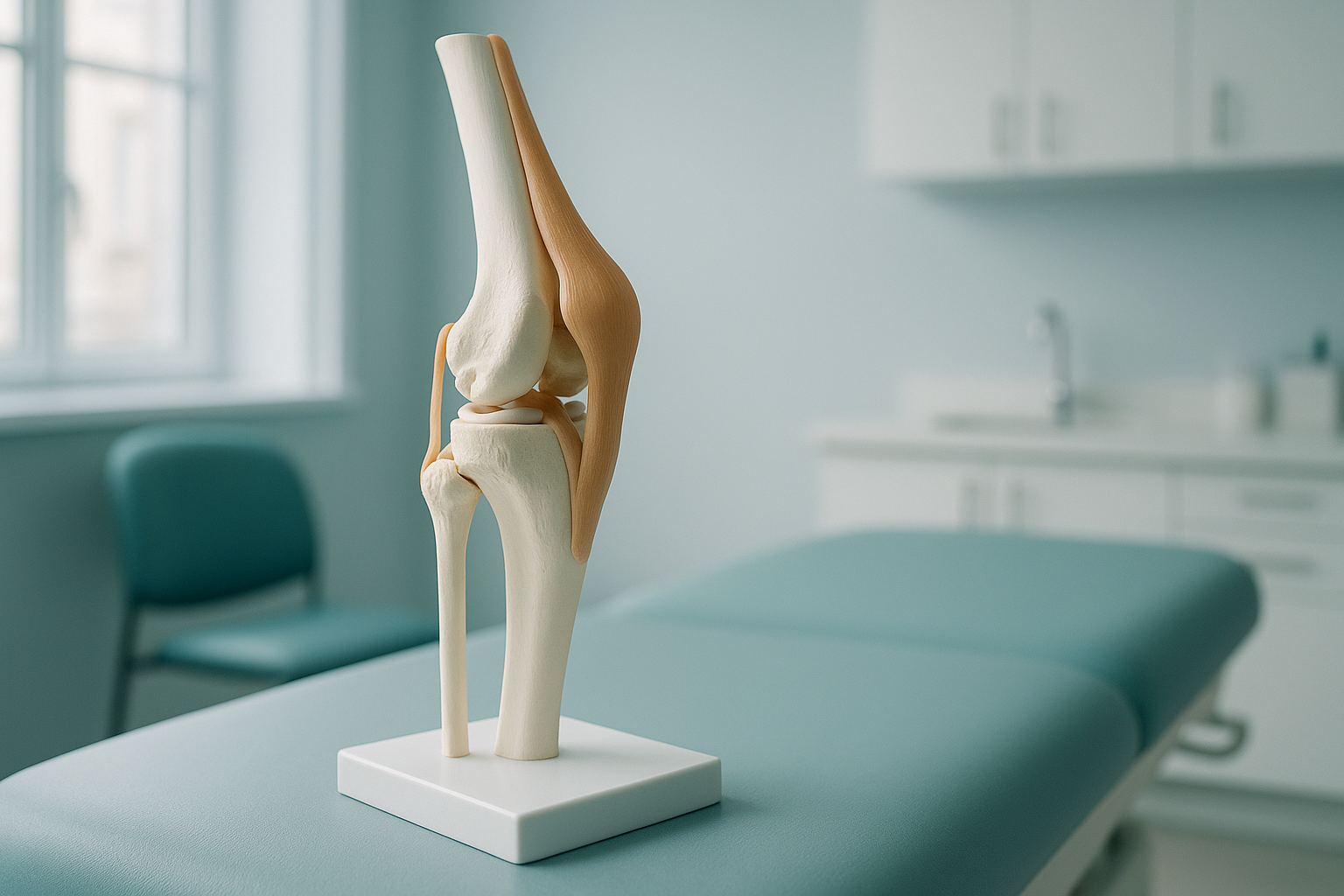
Your knee joint is where three bones meet: the thighbone (femur), the shinbone (tibia) and the kneecap (patella). Four main ligaments hold them together. Two types of cartilage cushion and protect the surfaces. Several tendons join your muscles to the bones, so you can bend, straighten and bear weight.
Key points
- The knee depends on three bones, four main ligaments, two types of cartilage and several tendons working together.
- Pain often shows up away from its true cause, because the whole leg shares the load.
- Scans taken lying down can miss alignment problems that only appear when you stand and bear weight.
- EOS standing imaging captures the full leg in one low-dose image, so your clinician can see how every part lines up under real conditions.
Knowing your knee anatomy helps you make sense of a diagnosis, ask better questions and choose the right next step. Below is a simple guide to every major part of the knee, what it does, and why alignment matters more than most people realise.
Which bones make up the knee joint?
Three bones form the knee joint: the femur (thighbone), the tibia (shinbone) and the patella (kneecap). A fourth bone, the fibula, sits beside the tibia on the outer side. It gives an anchor point for one of the ligaments, but it does not carry your body weight directly.
The longest and strongest bone in your body. Its rounded lower end rests on the tibia and glides when you bend or straighten the knee.
The main weight-bearing bone of the lower leg. The femur sits on its flat upper plateau, cushioned by cartilage.
A small, shield-shaped bone that sits inside the quadriceps tendon. It protects the front of the joint. It also acts as a lever, giving your thigh muscles more power when you straighten the knee.
Even a small shift in how these bones line up can change where force travels through the joint. That is why checking alignment matters, especially when a problem has been hard to explain.
What do the four main knee ligaments do?
Ligaments are tough bands of tissue that join bone to bone. The knee has four main ligaments. Each one stops a specific kind of unwanted movement.
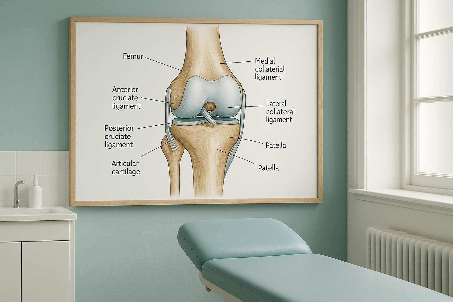
Anterior cruciate ligament. Stops the shinbone from sliding forwards under the thighbone and helps control rotation.
Posterior cruciate ligament. Stops the shinbone from sliding backwards. A PCL strain is less common than an ACL tear but can cause deep instability.
Medial collateral ligament. Resists forces pushing the knee inward. An MCL injury is one of the most common knee ligament problems.
Lateral collateral ligament. Resists forces pushing the knee outward. It runs along the outer side, joining the femur to the fibula.
When one ligament is weak or torn, the others work harder to make up for it. Over time, this extra load can cause cartilage wear and pain that seems unrelated to the first injury.
What does the cartilage in your knee do?
There are two types of cartilage inside the knee. Each has a different job.
Articular cartilage is the smooth, slippery coating on the ends of the femur, the top of the tibia and the underside of the patella. It lets the bones glide over each other with almost no friction. When this layer wears thin or softens, you might hear the term chondromalacia patella. This means damage on the underside of the kneecap.
Each knee has two menisci: the medial (inner) and the lateral (outer). These are crescent-shaped pads of tough, rubbery cartilage that sit between the femur and tibia. They act as shock absorbers, spread load evenly across the joint and help keep the femur stable on the tibia. A torn meniscus is one of the most common knee injuries.
Cartilage does not show up on a standard X-ray. But when standing imaging shows uneven joint spacing, it gives a strong clue that the cartilage on one side is thinner than on the other.
What are the main tendons around the knee?
Tendons join muscles to bones. Two tendons are especially important for how the knee works.
- Quadriceps tendon. Joins the four muscles at the front of your thigh to the top of the patella. It is the main driver when you straighten your knee.
- Patellar tendon. Runs from the bottom of the patella to the front of the tibia. When the quadriceps contracts, force passes through this tendon to straighten the lower leg. Overuse, or a kneecap that does not track well, can lead to patellar tendonitis.
Other tendons also play important roles in keeping the knee stable during movement. These include the hamstring tendons at the back and the iliotibial band on the outer side.
What are the joint capsule and synovium?
The joint capsule is a tough, fibrous sleeve that wraps around the whole knee. Inside that sleeve is a thin lining called the synovium (or synovial membrane). The synovium makes a small amount of synovial fluid, which lubricates and nourishes the cartilage. If the synovium becomes inflamed, it makes extra fluid and the knee swells. This is what people call "water on the knee".
When is MRI the better choice? EOS standing imaging shows bones, joint spacing and overall alignment very clearly. For soft-tissue detail, such as a suspected meniscus tear, ligament damage or an inflamed synovium, MRI is usually the better tool. Your clinician may recommend one or both, depending on the question they need to answer.
Why does whole-leg alignment matter for your knee?
Your knee does not work on its own. It sits between the hip and the ankle, and the alignment of the whole leg decides how force travels through the joint. If the hip tilts or the foot rolls inward, the knee is forced to make up for it. Over time, uneven loading wears one side of the cartilage faster and overloads certain ligaments or tendons.
An EOS standing scan captures your full leg, from hip to ankle, in a single weight-bearing image. Because you are upright and your muscles are working, the image shows how the bones and joints actually line up under real conditions. This often reveals misalignment that a lying-down scan would miss.
Ordinary X-rays, CT scans and MRI scans are usually taken while you lie down. In that position, gravity is removed and your muscles relax. The joint can look well aligned on the image, even when it shifts a lot under your body weight. That is why some knee problems are hard to explain with lying-down scans alone.
EOS imaging also uses about 90% less radiation than a standard X-ray, around ten times less. That makes it suitable when your clinician needs repeated follow-up images over time.
Before you start treatment, or change the one you are on, it is worth getting a detailed diagnosis. Knowing whether the cause sits in the pelvis, the legs or the spine changes what will actually help.
Want to see how your knee lines up under your own body weight? ScanAlign offers EOS standing imaging at 19 Harley Street, London, for patients travelling from abroad.
Book a free video consultationFrequently asked questions about knee anatomy
Three bones form the knee joint itself: the femur (thighbone), tibia (shinbone) and patella (kneecap). The fibula sits beside the tibia and anchors one ligament, but it is not a direct part of the joint surface.
A ligament joins bone to bone and keeps the joint stable. A tendon joins muscle to bone and passes on the force that moves your leg. Both are made of tough connective tissue, but they have different jobs.
Each meniscus is a rubbery, crescent-shaped pad that sits between the femur and tibia. The two menisci absorb shock, spread load across the joint and help keep the femur stable on the flat surface of the tibia.
Yes. Your body is a connected chain. If the hip tilts or the ankle rolls, the knee makes up for it to keep you upright. Over time, that extra stress can cause pain, cartilage wear or tendon overload at the knee, even though the root cause is elsewhere.
When you lie down, gravity is removed and your muscles relax. The joint can look well aligned on the image even though it shifts under your body weight. A standing scan shows the real picture. That is why alignment problems are sometimes only visible in a weight-bearing image.
Articular cartilage is the smooth, glassy coating on the ends of the femur, the top of the tibia and the underside of the patella. It lets the bones glide with almost no friction. When this layer wears thin, you may hear terms like osteoarthritis or chondromalacia.
EOS uses about 90% less radiation than a standard X-ray, around ten times less. This makes it a good option when several views or follow-up images are needed.
No. ScanAlign is a private, self-pay clinic. You can book a standing knee assessment yourself, without a referral from your doctor. Your images and report can then be shared with any clinician you choose.
EOS is designed for bones and alignment. For soft-tissue detail, such as a meniscus tear, ligament damage or nerve problems, MRI is usually the better tool. Your clinician may recommend one or both, depending on what they need to see.
The price for patients travelling from abroad is confirmed during the free video consultation, and more answers are on the ScanAlign FAQs page. The consultation comes first, so you can check whether the scan is right for you before committing.
See how your knee really lines up
A free video consultation helps you understand whether a standing scan is the right next step for your knee.
Book a free video consultationWritten by Abbas Dhami (Specialist Diagnostic Radiographer)
Sources
- NHS. "Knee pain." nhs.uk. Accessed 2024.
- American Academy of Orthopaedic Surgeons (AAOS). "Knee Ligament Injuries." orthoinfo.aaos.org. Accessed 2024.
- Drake RL, Vogl AW, Mitchell AWM. Gray's Anatomy for Students. 4th ed. Elsevier; 2019.
This page is general information and not a diagnosis.
ScanAlign is the trading name of CoreMed Solutions Ltd and operates under The Harley Street Hospital's CQC licence.