
Chondromalacia Patella: What Is It and How Is It Treated?
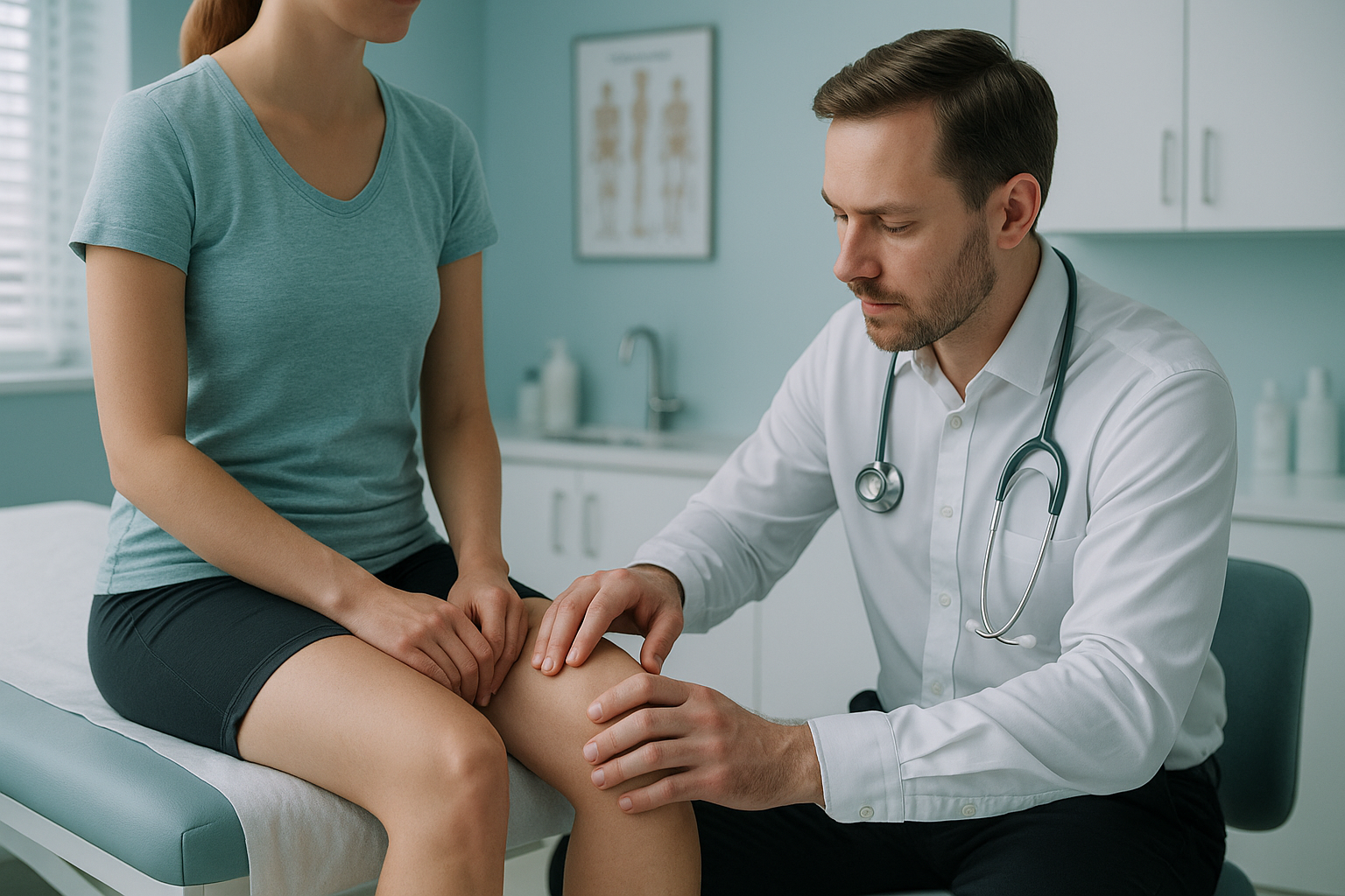
Chondromalacia patella is a softening or wearing of the cartilage on the underside of your kneecap. It causes pain at the front of the knee, especially on stairs, when squatting or after sitting for a long time. Treatment usually starts with targeted exercises and changes to your activity. But knowing why your kneecap tracks poorly is key to getting the right help.
Key points
- Chondromalacia means the cartilage behind your kneecap has softened, roughened or worn away.
- It is graded from 1 (mild softening) to 4 (exposed bone), but symptoms do not always match the grade.
- Poor kneecap tracking is often driven by alignment problems in the hip, leg or foot, not just the knee itself.
- Standing, weight-bearing imaging can show alignment problems that lying-down scans miss.
- Most people improve with the right exercises and guidance. Surgery is rarely the first option.
If you have been told you have chondromalacia patella, you probably have many questions. The name sounds alarming, but it simply describes damaged cartilage behind the kneecap. The real question is why it happened. That is where most standard tests fall short.
What does chondromalacia patella feel like?
Chondromalacia patella typically causes a dull, aching pain at the front of the knee. It tends to get worse with activities that put load on the kneecap. You may notice grinding or a gritty feeling when you bend and straighten your leg.
Important: if your knee suddenly locks or gives way, or swells a lot after an injury, see your doctor or go to the emergency department promptly. These symptoms may suggest a different problem that needs urgent assessment.
What causes cartilage softening behind the kneecap?
Cartilage softening behind the kneecap is usually caused by abnormal tracking. When the kneecap does not glide smoothly in its groove on the thighbone, some areas of cartilage take more load than they should. Over time, this uneven pressure breaks the cartilage down.
Several factors play a part. They are not always obvious from looking at the knee alone.
Your body is a connected chain from your feet to your head. A problem at one level can make another level compensate. This is why knee pain sometimes has its root cause in the hip or the foot. Treating only the knee, without understanding the bigger picture, can mean the problem keeps coming back.
What are the grades of chondromalacia patella?
Chondromalacia patella is graded from 1 to 4, based on how much the cartilage has changed. The grade is usually assessed on an MRI or during arthroscopy (a camera inside the knee). But your symptoms do not always match the grade. Some people with a higher grade have little pain, while others with a lower grade feel a lot of discomfort.
The grade alone does not decide your treatment plan. What matters more is your level of pain, your activity goals and, most importantly, whether an underlying alignment problem is driving the damage. Addressing the cause can slow further wear, even at higher grades.
Is chondromalacia the same as patellofemoral pain syndrome?
Not exactly, although the two conditions overlap a great deal. Patellofemoral pain syndrome (sometimes called runner's knee) is the broader term for pain around or behind the kneecap. Chondromalacia patella is a specific finding. It means the cartilage on the underside of the kneecap has actually softened or worn away. You can have patellofemoral pain without cartilage damage, but chondromalacia is one of its possible causes.
In practice, both conditions share the same underlying driver: abnormal kneecap tracking. That is why they are assessed in a similar way. Understanding your whole-leg alignment, from the hip down to the foot, helps make clear whether the cartilage damage is the source of your pain or a secondary finding.
How does a standing scan help with chondromalacia treatment?
A standing, weight-bearing scan shows your bones and joints under real-life load. This matters because lying-down imaging, such as a standard X-ray or MRI, relaxes your muscles. It can hide the very alignment problems that are pushing your kneecap off track. When you stand, gravity and your body weight show the true picture.
An EOS scan captures your full body, from head to feet, in a single standing image. It uses about 90% less radiation than a standard X-ray, around ten times less. This whole-body view lets a specialist measure the angles of your hip, knee and ankle together, and see exactly how they relate to one another.
For chondromalacia, this matters because the knee rarely works on its own. Your hip may rotate inward, your foot may roll in, or one leg may be slightly different in length. All of these change how your kneecap tracks. A scan of just the knee, taken lying down, can miss these links entirely.
If you have been managing knee pain for months without clear answers, a standing alignment assessment can help fill in the gaps.
Book a free video consultationWhen MRI is the better choice: EOS shows bones, joints and alignment clearly. For soft tissue detail, such as the cartilage surface itself, ligaments or nerves, MRI is usually the better tool. In many cases the two types of imaging complement each other. Together they give a fuller picture than either one alone.
What helps with chondromalacia patella?
Most people with chondromalacia improve with the right mix of exercises, changes to activity and understanding their alignment. Surgery is not usually the first step. It is typically kept for cases that have not responded to other approaches.
General management approaches
- Targeted strengthening. Exercises that build the muscles around the hip and thigh can improve how the kneecap tracks. A physiotherapist can tailor a programme to your specific needs.
- Changing your activity. Cutting back for a while on high-impact activities, like running or deep squats, can help settle symptoms while you build strength.
- Footwear and orthotics. If foot alignment is playing a part, supportive shoes or custom insoles may help correct the chain from below.
- Understanding your alignment. Knowing whether the problem is driven by your hip, your leg angle, your foot or a mix of these helps target your rehabilitation properly.
ScanAlign is a diagnostic imaging clinic. We do not provide treatment, but we can give you and your treating specialist the detailed alignment information that helps guide the right approach.
Before you start treatment, or change the one you are on, it is worth getting a detailed diagnosis. Knowing whether the cause sits in your pelvis, your legs or your spine changes what will actually help.
Learn more about how standing knee imaging works at ScanAlign.
Frequently asked questions about chondromalacia treatment
Mild chondromalacia (grade 1) can sometimes improve with rest, strengthening exercises and changes to activity. More advanced cartilage damage is unlikely to reverse fully. But symptoms can often be managed well by addressing the underlying alignment and muscle imbalances.
They are related but not the same. Chondromalacia specifically describes cartilage softening on the kneecap. If the cartilage loss progresses, it can develop into patellofemoral osteoarthritis. Finding and managing the tracking problem early may help slow that progression.
Exercises that strengthen the inner quadriceps, hip abductors and glutes are commonly recommended. Low-impact options, such as swimming or cycling on a higher seat, can also help. A physiotherapist should tailor any programme to your specific alignment and strength profile.
Surgery is usually considered only when structured rehabilitation has not improved symptoms after several months. Options can include arthroscopic smoothing of the cartilage or, in some cases, procedures to realign the kneecap. Your orthopaedic surgeon would discuss these if they are relevant to you.
No. ScanAlign is a private, self-pay clinic, so you can book directly without a referral from your doctor. Many people come to us after standard tests have not given them clear answers.
An EOS scan uses about 90% less radiation than a standard X-ray, around ten times less. This makes it one of the lowest-dose imaging options available for full-body skeletal assessment.
The price for patients travelling from abroad is confirmed during the free video consultation, and you can read more on the ScanAlign FAQs page.
EOS is a bone and joint imaging system. It does not image the cartilage surface directly. What it does show is the alignment of your whole leg under load. This shows the mechanical reasons your kneecap may be tracking abnormally. For direct cartilage assessment, MRI is typically used.
MRI is taken lying down, which relaxes your muscles and removes the effect of gravity. Alignment problems that only appear when you are standing and bearing weight can be invisible on a lying-down scan. A standing EOS assessment can sometimes show what an MRI misses.
You will have a short video call with a specialist. They will listen to your symptoms, review any imaging you already have, and advise whether a standing EOS scan is likely to add useful information for your case. There is no obligation to book a scan.
Still looking for answers about your knee pain?
A free video consultation can help you understand whether a standing alignment assessment in London is the right next step.
Book a free video consultation Learn about EOS imagingSources
- NHS. "Knee pain." nhs.uk, 2023. Overview of common causes of knee pain, including patellofemoral problems.
- Crossley KM, Stefanik JJ, Selfe J, et al. "2016 Patellofemoral pain consensus statement." British Journal of Sports Medicine, 2016;50:839-843.
- Illés T, Somoskeöy S. "The EOS imaging system and its uses in daily orthopaedic practice." International Orthopaedics, 2012;36(7):1325-1331.
Written by Abbas Dhami (Specialist Diagnostic Radiographer)
This page is general information and not a diagnosis.
ScanAlign is the trading name of CoreMed Solutions Ltd and operates under The Harley Street Hospital's CQC licence.