
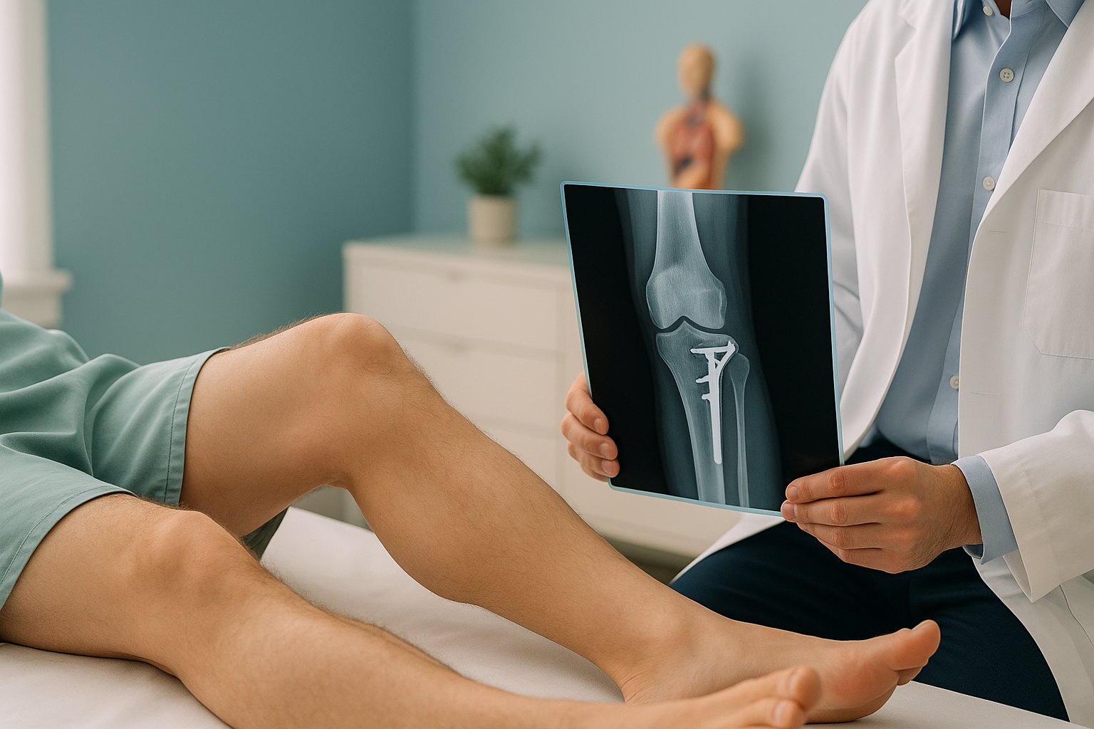
What Is a High Tibial Osteotomy, and Who Is It Suitable For?
A high tibial osteotomy is a knee realignment operation. It reshapes the top of the shinbone to move weight away from a worn inner part of the knee. It is designed for younger, active adults who want to keep their natural joint and delay or avoid a knee replacement. Precise standing imaging is essential for planning the correction.
Key takeaways
- High tibial osteotomy corrects bow-leg alignment to take load off the worn inner side of the knee.
- It is best suited to active adults with early to moderate arthritis in one compartment only.
- The surgery keeps your own joint, and may delay a knee replacement by many years.
- Accurate planning before surgery depends on a full-leg standing x-ray that maps the true weight-bearing line.
- EOS imaging uses about 90% less radiation than a standard x-ray, around ten times less.
What does a high tibial osteotomy do to the knee?
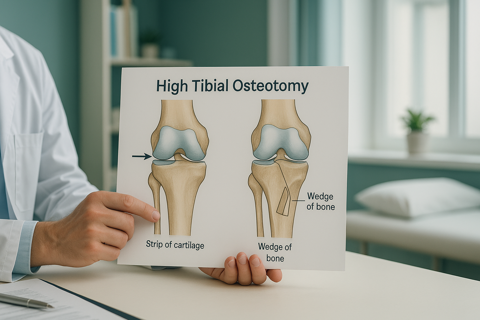
A high tibial osteotomy (often called HTO) is a planned, controlled cut through the upper part of the tibia, your shinbone. The surgeon opens or closes a small wedge of bone to change the angle of the leg. This moves the body's weight-bearing line from the damaged inner (medial) compartment of the knee to the healthier outer side.
Think of it like straightening an uneven table leg. When the leg is bowed inward, all your weight presses through one narrow strip of cartilage. Correcting the angle spreads the load more evenly. The goal is to preserve the joint. HTO does not remove the damaged surfaces and fit an artificial joint. It keeps your own knee intact and working.
HTO is most often used for varus malalignment, the medical term for bow legs. If your knees angle the other way (knock knees, or valgus alignment), a different procedure may be more suitable. You can read more about knock knee surgery separately.
Who is a good candidate for a high tibial osteotomy?
The ideal candidate is a younger, active adult with early to moderate arthritis limited to the inner compartment of the knee. HTO is seen as a joint-preserving option, so it works best when there is still healthy cartilage on the outer side.
- Active adults, usually under 60
- Bow-leg (varus) alignment
- Wear limited to the inner compartment
- Stable knee ligaments (or combined with ligament repair)
- Good range of movement in the knee
- Motivated to complete rehabilitation
- Widespread arthritis across the whole knee
- Severe cartilage loss on the outer side too
- Very limited knee bend (significant stiffness)
- Inflammatory arthritis such as rheumatoid disease
- Significant obesity that would overload the correction
Your surgeon will look at several factors before recommending HTO. Age alone does not rule you in or out. But the operation is generally aimed at people who are too young or too active for a knee replacement to be the best first option.
What are the types of high tibial osteotomy?
There are two main techniques. Both realign the leg, but they do it in different ways.
Opening wedge osteotomy is the more common technique today. The surgeon makes a cut on the inner side of the tibia and slowly opens a gap to correct the angle. A metal plate holds the bone in its new position while it heals. A bone graft or substitute may fill the gap.
Its advantages include precise control of the correction and the ability to adjust the angle during surgery. It also avoids disturbing the outer compartment of the knee.
Closing wedge osteotomy removes a small wedge of bone from the outer side of the tibia, then closes the gap to straighten the leg. This technique has a long track record. It heals quickly because the two bone surfaces are pressed together.
It is sometimes preferred when a large correction is needed or when the bone on the outer side is of better quality. However, it does change the height of the joint line slightly, which some surgeons prefer to avoid.
Your surgeon will choose the technique based on how much correction is needed, your bone quality, and whether any other procedures are being done at the same time.
What are the signs you might need a knee osteotomy?
Knee osteotomy is not usually the first thing your doctor considers. It usually comes up after simpler measures have not given enough relief. Common signs include:
If you also have sudden locking, giving way, numbness, weakness, or loss of bladder or bowel control, please get urgent medical help from your doctor or the nearest emergency department. These symptoms need prompt assessment rather than routine imaging.
Why does planning before surgery need standing imaging?
Accurate planning is the single most important factor in a successful high tibial osteotomy. The surgeon needs to know exactly where your weight-bearing line falls and how much correction to make. That information can only come from images taken while you are standing, with your skeleton carrying your weight as it does in daily life.
When you lie down for a standard x-ray or MRI, gravity is taken away. Your muscles relax, your joints decompress, and the true angle of your leg can shift. A full-leg standing x-ray captures the whole limb from hip to ankle in one image, under full body weight. This shows the real mechanical axis your surgeon needs to plan around.
At ScanAlign, private standing imaging, EOS technology captures this full-length standing view with about 90% less radiation than a standard x-ray, around ten times less. It produces a single, calibrated image of both legs at the same time, so the surgical team can compare sides and plan the correction with confidence.
Considering HTO surgery? Your surgeon may need standing alignment imaging before finalising the surgical plan. We can arrange this quickly at 19 Harley Street, London, and your images can be shared directly with your surgical team.
Book a free video consultationWhen MRI is the better tool: EOS shows bones, joints and alignment clearly. If your surgeon also needs to check soft tissue, cartilage, meniscus tears or ligament damage, MRI is usually the better choice. Many patients being planned for HTO will need both types of imaging.
What does recovery from a tibial osteotomy look like?
Recovery from a high tibial osteotomy is gradual. The bone needs time to heal in its new position, and the muscles around the knee need rebuilding. Everyone recovers at a different pace, but here is a general outline of what to expect.
You will likely use crutches and may wear a brace. Putting weight on the leg is usually limited at first, then increased step by step under your physiotherapist's guidance.
The bone continues to heal. Most people can start putting more weight through the leg and stop using crutches. Gentle exercises help bring back range of movement.
Strengthening work increases. Many people return to low-impact activities, office work and driving during this period.
Return to sport and higher-impact activities is often possible, depending on the sport and your surgeon's guidance. Full recovery can take up to a year.
Follow-up imaging after the osteotomy can confirm that the bone has healed and the alignment is where the surgeon intended. Standing x-rays are especially useful for checking the corrected mechanical axis once you are fully putting weight on the leg again.
Before you start treatment, or change the one you are on, it is worth getting a detailed diagnosis. Knowing whether the cause sits in the pelvis, the legs or the spine changes what will actually help.
Frequently asked questions about high tibial osteotomy
Neither is better for everyone. HTO keeps your own joint and is aimed at younger, active people with wear on one side of the knee. A knee replacement is more suitable when arthritis affects the whole joint. Your surgeon will help decide which is right for you.
Results vary. Many studies report the benefits lasting ten years or longer, especially when patient selection and alignment correction are accurate. Some people may need a knee replacement later in life, but HTO can significantly delay that step.
Many patients do return to sport, including running, cycling and skiing. The timeline depends on healing, your rehabilitation and your surgeon's advice. Return to high-impact activities is usually considered from around nine to twelve months.
In an opening wedge, the surgeon cuts the bone and opens a gap on the inner side to correct the angle. In a closing wedge, a small piece of bone is removed from the outer side and the gap is closed. Both aim to straighten the leg and spread the load differently.
A full-leg standing image shows the mechanical axis from hip to ankle under real body weight. This is the only reliable way to measure the exact angle of malalignment and plan how much correction is needed. Lying-down images can underestimate the true deformity.
Yes. EOS uses about 90% less radiation than a standard x-ray, around ten times less. It is a well-established system used in hospitals worldwide. You can read more about safety and what to expect on our FAQs page.
No. You can refer yourself as a private patient. If your surgeon or physiotherapist would like to refer you directly, they can refer a patient for imaging through our clinician registration page.
The price for patients travelling from abroad is confirmed during the free video consultation.
HTO is mainly designed for varus (bow-leg) malalignment. If you have valgus (knock-knee) alignment, a different type of osteotomy around the knee may be more suitable. Read more about knock knee surgery for details on that correction.
Yes, certainly. Many patients and surgical teams arrange standing alignment imaging ahead of the operation, so the plan is finalised well in advance. You can book pre-operative alignment imaging through a free video consultation to get started.
Need standing alignment imaging for your knee?
Whether you are looking into a high tibial osteotomy or your surgeon has asked for planning images before surgery, we can help.
Book a free video consultationWritten by Abbas Dhami (Specialist Diagnostic Radiographer)
Sources
- Brinkman J-M, Lobenhoffer P, Agneskirchner JD, et al. "Osteotomies around the knee: patient selection, stability of fixation and bone healing in high tibial osteotomies." The Journal of Bone & Joint Surgery (British Volume), 2008.
- W-Dahl A, Toksvig-Larsen S, Roos EM. "Association between knee alignment and knee pain in patients surgically treated with high tibial osteotomy." Knee Surgery, Sports Traumatology, Arthroscopy, 2009.
- National Institute for Health and Care Excellence (NICE). "Osteoarthritis: care and management." Clinical guideline CG177, updated 2022.
This page is general information and not a diagnosis.
ScanAlign is the trading name of CoreMed Solutions Ltd and operates under The Harley Street Hospital's CQC licence.