

What Does a ScanAlign Report Include?
A ScanAlign report has three parts. First, calibrated standing images. Second, a structured table of measurements for the spine, pelvis and legs. Third, a written commentary from a radiologist. The images come in DICOM format along with an annotated PDF. Together, they give you everything you need to plan treatment with confidence.
Key points
- Full-length standing images taken on the EOS system, with about 90% less radiation than a standard X-ray (around ten times less).
- A structured table of alignment measurements for the spine, pelvis and legs.
- Written clinical commentary from a consultant radiologist.
- Images sent in DICOM and annotated PDF formats.
- A report made for direct clinical use by surgeons, physiotherapists and other referring professionals.
When you refer a patient to ScanAlign, standing EOS imaging, you get a report built to support clinical decisions, not a general imaging summary. This applies whether your patient travels to London from Pakistan or anywhere else. This page goes through each section of the report, so you know exactly what to expect.
What sections are in an imaging report?
Every ScanAlign report has three main sections: images, measurements and clinical commentary. Together, they give a complete picture of how the patient's body is aligned when standing, from head to feet.
Written findings from a consultant radiologist, covering the main observations and any abnormalities.
Structured, calibrated values for spinal curves, pelvic parameters and lower-limb alignment.
Full-length standing AP and lateral images in DICOM format, plus an annotated PDF for quick reference.
What measurements are in a spine and alignment report?
The measurement table is the clinical core of the report. The EOS system takes standing, weight-bearing images at true 1:1 scale. So every value reflects how the body is loaded in real life. Measurements are grouped by body region.
Spinal parameters
The report covers coronal and sagittal alignment of the whole spine. This includes Cobb angle measurement for any coronal curves. It also includes sagittal values such as thoracic kyphosis, lumbar lordosis and the sagittal vertical axis (SVA). All values are worked out from the standing image. So they reflect the patient's true posture under gravity.
Pelvic parameters
Pelvic incidence, pelvic tilt and sacral slope are reported together as a set. These three values depend on each other and are essential for planning sagittal balance. Any leg-length discrepancy is also measured here.
Lower-limb alignment
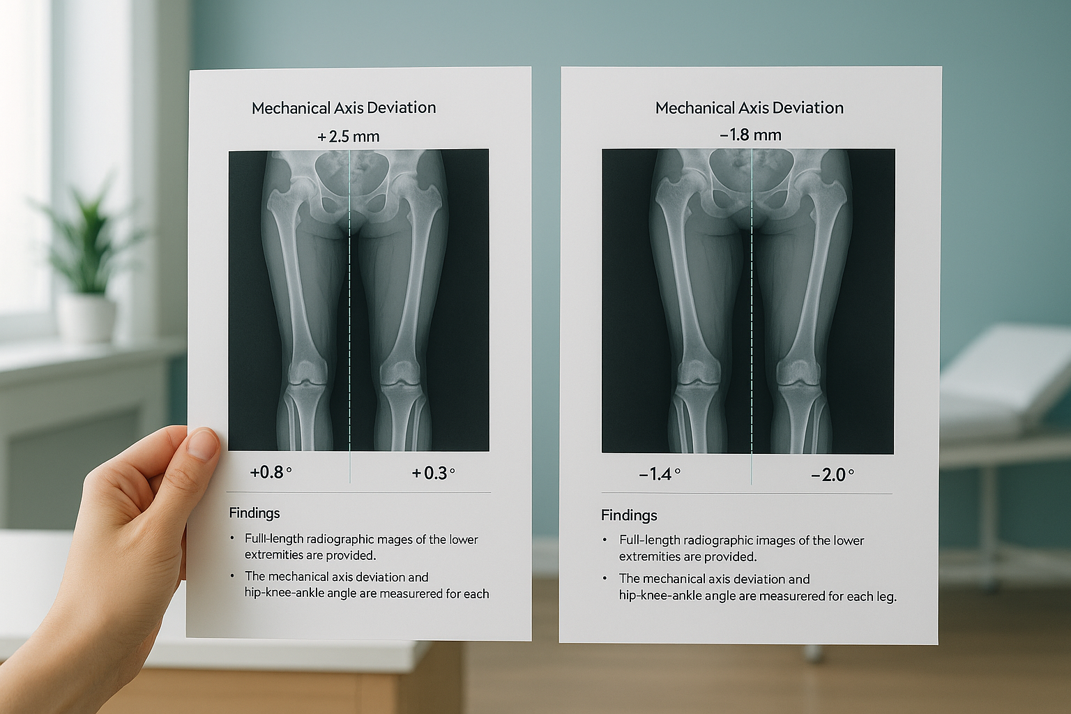
For patients scanned with a full-length protocol, the report includes mechanical axis deviation and the hip-knee-ankle (HKA) angle for each leg. This is especially useful for planning before knee or hip arthroplasty.
What image formats come with the report?
Images are sent in two formats. DICOM files can be opened in your PACS or any standard viewing software for detailed analysis. An annotated PDF is also included. It shows the key measurements laid over the images. This makes it easy to discuss findings with patients or colleagues without specialist software.
The EOS system uses slot-scanning technology. So the images are taken at true 1:1 scale with minimal distortion. This means you can trust the measurements without adjusting for magnification.
What does a sample EOS scan report look like?
Below is an anonymised example of the measurement table you would receive. The actual report also includes the full radiologist commentary and the images. For an explanation written for patients, see how EOS scan results are reported or read more about what an EOS scan report shows.
| Parameter | Region | Example value |
|---|---|---|
| Thoracic kyphosis (T4-T12) | Spine | 38° |
| Lumbar lordosis (L1-S1) | Spine | 52° |
| Coronal Cobb angle | Spine | 12° (T6-L1 convex left) |
| Sagittal vertical axis | Spine | +18 mm |
| Pelvic incidence | Pelvis | 54° |
| Pelvic tilt | Pelvis | 14° |
| Sacral slope | Pelvis | 40° |
| Leg-length discrepancy | Pelvis / limbs | 7 mm (left shorter) |
| Mechanical axis, left | Lower limb | 4 mm medial |
| HKA angle, right | Lower limb | 178° |
These values are for illustration only and do not belong to a real patient.
Why does a standing report show more than a lying-down scan?
When a patient lies down for a regular X-ray, CT or MRI, gravity is taken away and the muscles relax. Curves may flatten, a leg-length difference is no longer loaded, and pelvic tilt shifts. The result is a snapshot that does not match how the patient really stands and moves.
EOS scans the patient upright and bearing weight. So every measurement in the report reflects the real forces acting on the skeleton. For surgical planning, especially fusion levels, osteotomy correction or joint replacement alignment, this difference can change the plan completely.
The body is a connected chain from feet to head. A tilt at the pelvis can force the spine above it to compensate, and pain often shows up away from the true cause. A standing whole-body report shows these compensations in a way that lying-down imaging of a single region simply cannot.
Weight-bearing, full-length images. Gravity loads the joints and spine in the normal way. Measurements reflect the patient's true posture. You can see how one region compensates for another. 1:1 scale with no magnification error.
Unloaded images of separate segments. Muscles relax, curves may reduce, and the position of the pelvis shifts. Views of a single region can miss how one area affects another. Magnification changes with the distance between the patient and the detector.
Does the report cover soft-tissue findings?
EOS is designed to show bones, joints and alignment with exceptional clarity at very low radiation. For soft-tissue assessment, including discs, nerves, ligaments or cartilage, MRI is usually the better tool. The two work well together. Many clinicians use EOS for alignment planning and MRI for diagnosis at the tissue level.
Before your patient starts treatment, or changes the treatment they are on, a detailed diagnosis is worth getting. Knowing whether the cause sits in the pelvis, the legs or the spine changes what will actually help.
A note on MRI: EOS is excellent at showing skeletal alignment under load. For soft tissue, discs or nerves, MRI is usually the better choice. If your clinical question is mainly about soft tissue, MRI may be more suitable as the first test.
Would you like to see a full sample report or discuss a particular case? We are happy to take you through the format in a short call.
Book a free video consultationFrequently asked questions
Every report includes calibrated standing images (DICOM and annotated PDF), a structured table of measurements for the spine, pelvis and legs, and written commentary from a consultant radiologist.
Images come in standard DICOM format for loading into your PACS, along with an annotated PDF for quick review or for discussion with the patient.
The measurement table includes spinal values (thoracic kyphosis, lumbar lordosis, Cobb angles, SVA), pelvic parameters (pelvic incidence, pelvic tilt, sacral slope, leg-length discrepancy) and lower-limb alignment (mechanical axis, HKA angle), as clinically relevant.
Every report is reviewed and written by a consultant radiologist with experience in musculoskeletal imaging. The commentary highlights the key findings and any abnormalities relevant to the clinical question.
Yes. When you refer a patient, you can state the clinical question and any extra measurements you need. The protocol is adjusted to match.
The EOS system uses about 90% less radiation than a standard X-ray, around ten times less. This makes it especially suitable for patients who need repeated imaging or full-length views.
No. Patients can refer themselves, and clinicians can refer directly. You can start by contacting ScanAlign to discuss the case, or complete a referral online.
Yes. Standing, weight-bearing images at true 1:1 scale are widely used for planning spinal fusion levels, osteotomy corrections and joint replacement alignment. The calibrated measurements are ready for direct use in surgical planning software.
Yes. Contact ScanAlign directly and we can share an anonymised sample report. You can then judge the format and level of detail before referring your first patient.
For patients travelling from abroad, the price is confirmed during the free video consultation. You can also visit the FAQs and cost page.
Ready to refer a patient or look at the report format?
Book a short call to discuss your clinical question, or refer a patient directly.
Book a free video consultationSources
- Dubousset, J. et al. "EOS: A New Imaging Concept." European Spine Journal, 2010. Overview of EOS low-dose biplanar imaging and its clinical uses.
- The Royal College of Radiologists. Standards for the Reporting of Imaging Investigations. Guidance on structured radiology report content in the UK.
- Lazennec, J.Y. et al. "Pelvis and Total Hip Arthroplasty Acetabular Component Orientations in Sitting and Standing Positions." Orthopaedics & Traumatology: Surgery & Research, 2011. Shows how pelvic parameters change between lying and standing.
Written by Abbas Dhami (Specialist Diagnostic Radiographer).
This page is general information and not a diagnosis.
ScanAlign is the trading name of CoreMed Solutions Ltd and operates under The Harley Street Hospital's CQC licence.