
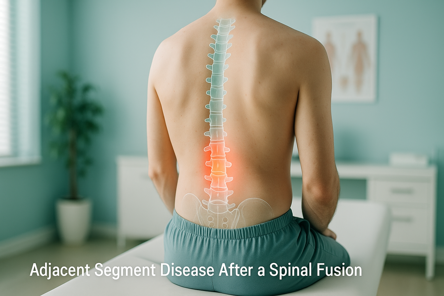
What Is Adjacent Segment Disease After a Spinal Fusion?
Adjacent segment disease is new pain, stiffness or instability in the spinal levels directly above or below an earlier fusion. The fused section no longer moves. So the discs and joints next to it take on extra load. Over months or years, this can speed up wear and cause symptoms in a part of the spine that was healthy before.
Key points
- Adjacent segment disease affects the levels next to a spinal fusion, not the fusion itself.
- It is driven by changed load and movement, and may take years to appear.
- Overall spinal balance matters. A fusion that changes your posture can speed up wear elsewhere.
- Standing, weight-bearing imaging shows alignment under real load. Lying-down scans can miss this.
- Early monitoring helps your surgeon plan the right next step before symptoms get worse.
Spinal fusion is a well-established operation. It stabilises a painful or unstable segment and, for many people, gives lasting relief. But the spine is a chain of connected segments. When one link is locked in place, the links above and below have to work harder. Adjacent segment disease describes what happens when that extra demand causes real problems.
This is not a criticism of the original surgery. Fusion does its job. The challenge is that the rest of the spine still needs to move, bend and absorb shock. The segments closest to the fusion carry the biggest share of that burden.
What does adjacent segment disease feel like?
The most common symptom is new or different pain. It appears months or years after a fusion that at first felt successful. The pain usually sits above or below the fused area, not in it.
Urgent note. If you develop sudden numbness or weakness in your legs, or any loss of bladder or bowel control, contact your doctor or go to the nearest emergency department immediately. These symptoms need urgent medical attention, not a routine scan.
Why does pain develop above or below a spinal fusion?
Extra mechanical stress on the neighbouring segments is the main driver. Several factors work together.
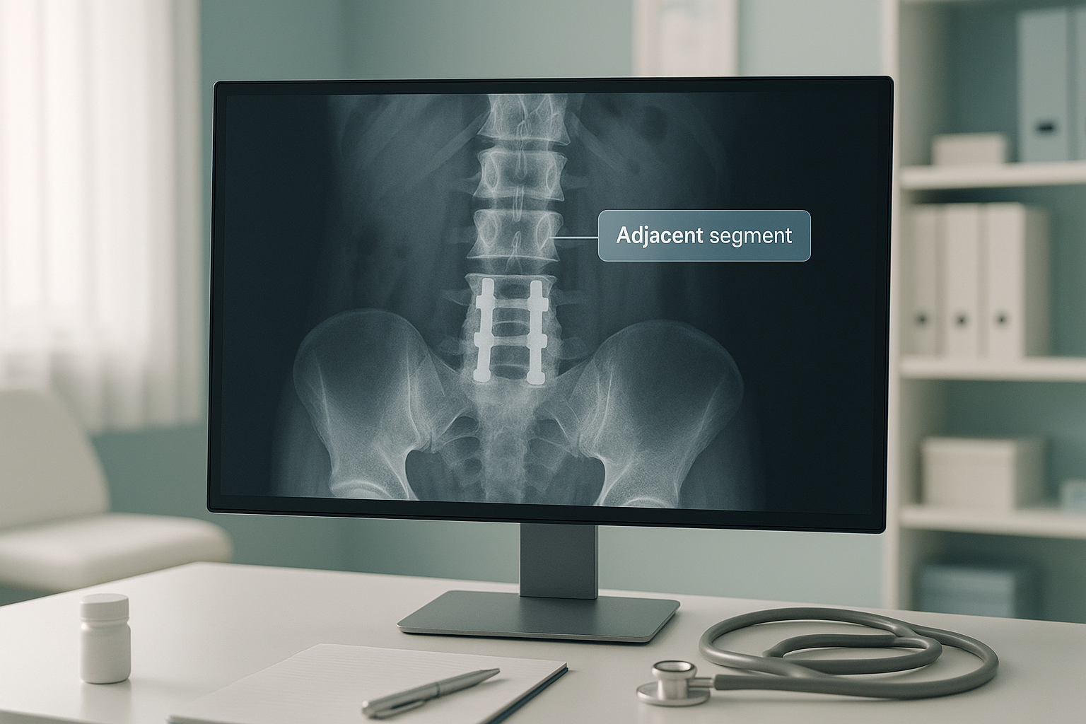
Sometimes a related issue is retrolisthesis, where a vertebra slips backward at an adjacent level. This is one of the structural changes that can develop when load is shared out differently after fusion.
How does a standing scan help after spinal fusion?
A standing, weight-bearing scan shows your spine under real load, exactly the way it behaves when you walk, stand and feel pain. Lying-down imaging relaxes your muscles and lets gravity settle things into a resting position. This can hide alignment shifts that only appear upright.
You stand naturally inside the scanner. Gravity pulls on your spine the way it does all day. This shows how your fusion, your adjacent levels and your overall posture relate to each other. ScanAlign, unstitched full-body standing imaging captures you from head to feet in a single pass. Your specialist can then see whether the fusion has shifted your balance elsewhere.
When you lie flat for a conventional X-ray, CT or MRI, the disc spaces open up and small slips may reduce. The natural adjustments your body makes through the day disappear. The result can look more reassuring than it really is. Key alignment problems may not show at all.
For adjacent segment disease, standing imaging is especially useful because it shows whether your spinal curves have changed since the fusion. A loss of the lower back's natural inward curve, sometimes called flatback syndrome, can force your body to make up for it by bending the hips and knees. This creates a forward-leaning posture. These shifts are invisible on a scan taken lying down.
EOS imaging uses about 90% less radiation than a standard X-ray, around ten times less. For someone who has already had many scans before and after surgery, that lower dose is a meaningful advantage for ongoing monitoring.
When MRI is the better tool. EOS shows bones, joints and alignment beautifully. But for soft-tissue detail, such as disc herniations, nerve compression or spinal cord changes at an adjacent level, MRI is usually the better choice. Your surgeon may want both: a standing alignment image and a detailed MRI of the affected discs.
What can you do about adjacent segment disease?
The right next step depends on how severe your symptoms are, what the imaging shows and your surgeon's assessment. ScanAlign is a diagnostic imaging clinic, not a treatment provider. Our role is to give you and your clinical team the clearest possible picture of what is happening in your spine.
Common approaches your specialist may discuss include:
- Monitoring. If changes are early and symptoms are mild, regular standing scans can track progress over time without rushing into more surgery.
- Physiotherapy and rehabilitation. Targeted strengthening around the adjacent segments can help manage symptoms and slow further wear.
- Injections or pain management. These can help control symptoms while decisions about longer-term care are made.
- Revision or extension surgery. When degeneration at the adjacent level becomes severe, extending the fusion or doing a further procedure may be considered. Accurate alignment data from a standing scan helps your surgeon plan this carefully.
Had a spinal fusion and noticing new pain? A standing spine assessment can show your surgeon exactly what is happening at the adjacent levels.
Book a free video consultationIs a standing spine assessment right for you?
A standing scan is particularly useful for people in certain situations. It is not always the right first step for everyone.
- Had a spinal fusion and now have new or worsening pain
- Feel your posture has changed since surgery
- Have been told you have degeneration at the adjacent level
- Are considering revision surgery and need alignment data
- Want to monitor your spine over time with low-dose imaging
- Have never had spinal surgery and suspect a disc or nerve problem (MRI first)
- Cannot stand without support for a few seconds
- Have sudden severe symptoms that need urgent care
Before you start a treatment, or change the one you are on, it is worth getting a detailed diagnosis. Knowing whether the cause sits in the pelvis, the legs or the spine changes what will actually help.
Frequently asked questions
It is new degeneration, pain or instability in the spinal segments directly above or below an earlier fusion. The fused section no longer moves, so the neighbouring levels take on extra load and can wear out faster.
Changes at the adjacent level that show on imaging are common and do not always cause symptoms. Clinically significant adjacent segment disease, meaning changes that cause new pain or disability, affects a smaller share of fusion patients over time. The likelihood increases with longer follow-up.
It can develop any time from a year or two after surgery to well over ten years later. There is no fixed timeline, which is why ongoing monitoring can be valuable.
No. Many people live comfortably after fusion without developing significant adjacent segment disease. The risk depends on factors like the number of levels fused, overall spinal balance and general disc health.
When you stand, gravity loads your spine the same way it does all day. Alignment shifts, balance changes and adjustments that are hidden when you lie flat become visible. This gives your surgeon a more accurate picture of what your spine is actually doing.
Yes. EOS uses X-ray technology, not magnetic fields, so metal implants are not a concern. The system also produces less scatter around metalwork than conventional X-rays, often giving a clearer image near the hardware.
EOS delivers about 90% less radiation than a standard X-ray, around ten times less. For patients who need repeated imaging to monitor their spine after surgery, this lower dose is a real benefit.
No. You can refer yourself. Many people come to ScanAlign on their own, and we provide imaging that they can share with their surgeon or consultant afterwards.
The price for patients travelling from abroad is confirmed during the free video consultation, which you can arrange on our booking page.
The scan provides detailed alignment and structural data. Whether a second surgery is needed is a decision for your surgeon. But the imaging gives them the objective information they need to make that recommendation with confidence.
Worried about pain after your spinal fusion?
A free video consultation lets you discuss your situation with a specialist radiographer from home, before you plan any trip to London. There is no obligation to book a scan.
Book a free video consultationAuthor: Abbas Dhami (Specialist Diagnostic Radiographer)
Sources
- Helgeson MD, Bevevino AJ, Hilibrand AS. Update on the evidence for adjacent segment degeneration and disease. The Spine Journal. 2013;13(3):342-351.
- Harrop JS, Youssef JA, Maltenfort M et al. Lumbar adjacent segment degeneration and disease after arthrodesis and total disc arthroplasty. Spine. 2008;33(15):1701-1707.
- National Institute for Health and Care Excellence (NICE). Low back pain and sciatica in over 16s: assessment and management. NICE guideline NG59 (updated 2020).
This page is general information and not a diagnosis.
ScanAlign is the trading name of CoreMed Solutions Ltd and operates under The Harley Street Hospital's CQC licence.