
What Is a Vertebral Compression Fracture, and Why Am I Losing Height?
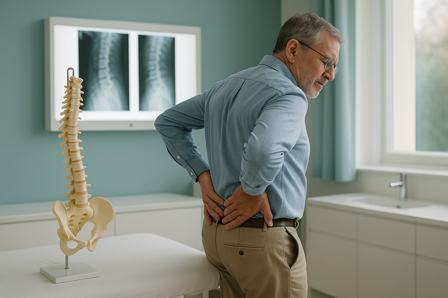
A vertebral compression fracture happens when a bone in your spine collapses under load. This is often caused by osteoporosis. The front of the vertebra crumbles into a wedge shape. This shortens your spine, and you lose height. Many people never feel a dramatic break. Instead, they notice their back slowly rounding, back pain, or their clothes fitting differently.
Key takeaways
- Compression fractures are the most common fracture linked to osteoporosis, especially after age 50.
- Even one collapsed vertebra can make you noticeably shorter. More than one fracture speeds up the change.
- Pain may be sudden and sharp, or it may build slowly over weeks.
- A standing scan shows how a fracture changes your spinal alignment under real weight-bearing load.
- If you have sudden severe back pain, numbness, weakness, or loss of bladder or bowel control, get urgent medical help from your doctor or your nearest emergency department.
What are the symptoms of a spinal compression fracture?
The most common sign is a sudden, sharp pain in the middle or lower back. It gets worse when you stand or walk, and eases when you lie down. But not everyone has a dramatic moment of pain. Some fractures happen slowly. The first clue may be that you are getting shorter, your posture is rounding forward, or your belt sits higher than it used to.
Seek urgent help. If you develop sudden severe back pain along with numbness or weakness in your legs, or any change in bladder or bowel control, contact your doctor or go to your nearest emergency department. These symptoms need immediate medical assessment.
What causes a vertebral compression fracture?
The overwhelming majority of compression fractures are caused by osteoporosis, a condition where bones slowly lose density and become fragile. In healthy bone, a vertebra can handle everyday loads. In weakened bone, something as minor as stepping off a kerb, a forceful sneeze, or bending forward to pick up a bag can be enough. The front edge of the vertebra collapses into a wedge shape.
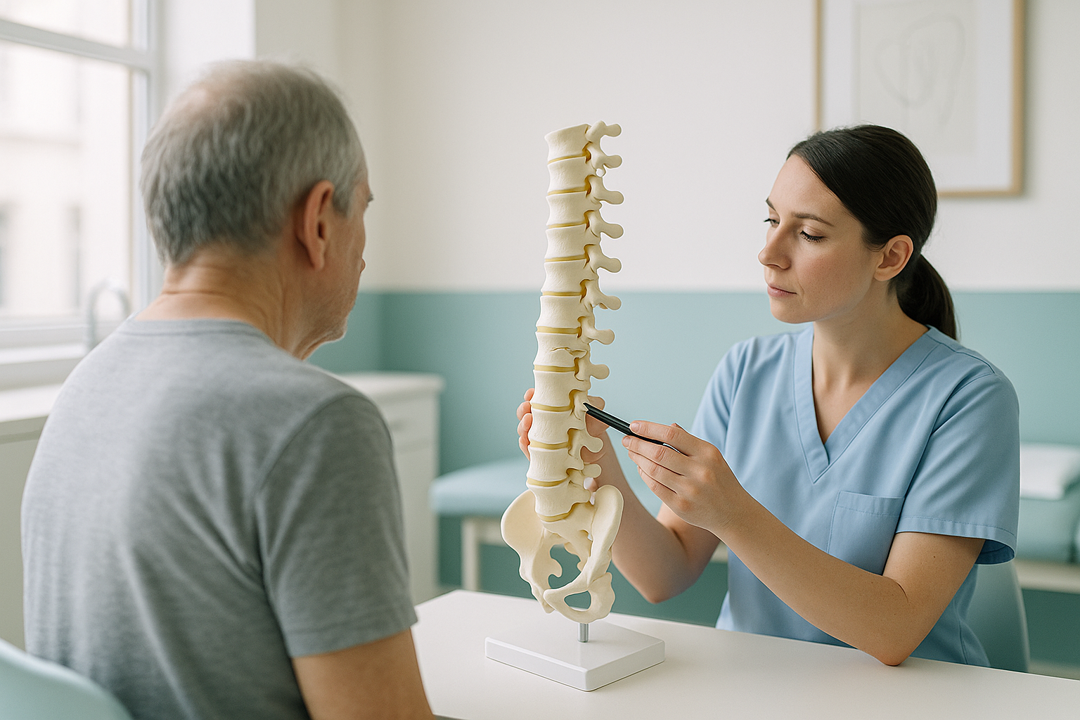
When the front of a vertebra collapses more than the back, the bone takes on a wedge shape. Stack several wedge-shaped vertebrae on top of each other and the spine curves forward. Over time, that forward curve is what takes away your height and changes your posture.
Why does a standing scan show compression fractures differently?
A standing scan captures your spine under real gravity. This is exactly when a compression fracture has its greatest effect. When you lie down for a standard X-ray, CT or MRI, gravity is removed. The fractured vertebra is partly unloaded. The full extent of the collapse, and its knock-on effects, can be hidden.
A standing side view shows how the fractured vertebra behaves under your full body weight. It reveals the true amount of collapse at the front of the bone, any forward tilt of the spine, and how the rest of your body compensates, from the pelvis tilting to the knees bending. This gives your specialist a complete picture of your sagittal balance.
Lying down relaxes your muscles and removes gravity. The fractured vertebra may partly decompress, so the collapse looks less severe than it really is. The image also cannot show how the rest of your body adapts when you are upright. That is the position in which you actually feel your symptoms.
Your body is a connected chain from your feet to your head. When one vertebra collapses, the spine tilts forward. Your pelvis, hips and knees all adjust to keep you upright. That compensation can cause pain in areas far from the fracture itself. ScanAlign standing EOS imaging captures the full chain in a single standing image, using about 90% less radiation than a standard X-ray, around ten times less.
When MRI is the better choice: EOS shows bones, joints and alignment. If your specialist needs to look at the spinal cord, nerves, discs or soft tissue around a fracture, MRI is usually the better tool. Both types of imaging can work together to give a complete picture.
What happens after a compression fracture is found?
ScanAlign is a diagnostic imaging clinic, not a treatment centre. Imaging gives you and your specialist a clear, accurate picture of what has happened to your spine. This helps plan the right next steps. Those steps depend on how severe the fractures are and how many there are.
For many people, care involves pain relief, changes to activity, and sometimes a brace while the bone heals. Where fractures are more severe, or are not responding to non-surgical care, procedures such as kyphoplasty or vertebroplasty may be discussed. Your treating clinician will also want to address your underlying bone health, to reduce the risk of more fractures.
Accurate standing alignment imaging is especially useful here. It shows whether the fracture has changed your overall spinal balance, and by how much. This helps your specialist decide which approach suits you best.
Worried about height loss or a possible compression fracture? A standing EOS scan in London shows the full picture.
Book a free video consultationIs a standing spine scan right for me?
A standing EOS scan is most helpful when you or your clinician want to understand how a fracture, or a suspected fracture, affects your whole spinal alignment under real load.
Before you start treatment, or change the treatment you are on, it is worth getting a detailed diagnosis. Knowing whether the cause sits in the pelvis, the legs or the spine changes what will actually help.
- Have been told you have a compression fracture and want to see its effect on your posture
- Are losing height and want to know why
- Have osteoporosis and back pain that keeps coming back
- Need imaging before or after a spinal procedure
- Have sudden leg weakness, numbness, or bladder/bowel changes (seek urgent medical help)
- Need soft-tissue or nerve detail (MRI is usually better)
- Have a suspected fracture from high-energy trauma that needs emergency assessment
Frequently asked questions about compression fractures
Many compression fractures do heal with time, rest and proper pain management. The bone knits back together, but the wedge shape usually remains. This means the height loss and any change in posture are typically permanent, even after healing.
A single fracture might reduce your height by a small amount. Several fractures, especially across several vertebrae, can together cause several centimetres of height loss and a visible forward curve.
It can feel like a sudden sharp pain in the middle or lower back, often set off by bending or lifting. Some people describe it as a deep ache that gets worse with standing and eases when lying flat. Others have no obvious pain at all and only notice changes in posture.
No. ScanAlign accepts self-referrals. You can book a standing spine scan directly through a free video consultation, without a letter from your doctor or specialist.
Yes. An EOS scan uses about 90% less radiation than a standard X-ray, around ten times less. You simply stand still for a few seconds. There is no injection, no enclosed space, and it is very gentle on the body.
Yes. When several vertebrae in the thoracic spine collapse into wedge shapes, the combined effect pushes the upper back into a rounded curve. Over time this can develop into what is commonly called a dowager's hump, known medically as kyphosis.
Lying down removes gravity. The fractured vertebra is partly unloaded, which can make the collapse look less severe. A standing image shows the fracture under your real body weight. It reveals how the fracture affects your overall spinal alignment and balance.
Both are minimally invasive procedures used to stabilise a fractured vertebra. They differ in technique and in when they are suitable. You can read a detailed comparison in our kyphoplasty or vertebroplasty guide.
ScanAlign is a private, self-pay clinic. The price for patients travelling from abroad is confirmed during the free video consultation, where you can also discuss whether a scan is right for you. The consultation is free and comes with no obligation.
Yes. A collapsed vertebra shifts your centre of gravity forward. Your body compensates by tilting the pelvis, bending the knees, or tightening muscles elsewhere. This changes your sagittal balance and can make walking feel less steady or more tiring.
See your spine the way it actually stands
A free video consultation takes a few minutes, and you can join it from home. We will help you understand whether a standing EOS scan at 19 Harley Street, London is the right next step for you.
Book a free video consultationWritten by Abbas Dhami (Specialist Diagnostic Radiographer)
Sources
- NHS. "Osteoporosis." nhs.uk. Accessed 2024.
- Royal Osteoporosis Society. "Vertebral Fractures." theros.org.uk. Accessed 2024.
- Old JL, Calvert M. "Vertebral compression fractures in the elderly." American Family Physician. 2004;69(1):111-116.
This page is general information and not a diagnosis.
ScanAlign is the trading name of CoreMed Solutions Ltd and operates under The Harley Street Hospital's CQC licence.