
What Is a Trendelenburg Gait, and What Does It Say About Your Hip?
A Trendelenburg gait is a way of walking where one side of the pelvis drops each time you step onto the other leg. It usually means the hip abductor muscles on the standing leg are weak or not working properly. It is a clinical sign, not a diagnosis in itself. Finding the cause behind it often needs imaging of the whole pelvis and legs while you are bearing weight.
Key points
- A Trendelenburg gait happens when the hip abductor muscles cannot hold the pelvis level while you stand on one leg.
- Common causes include gluteus medius weakness, hip joint problems, nerve problems and a difference in leg length.
- Imaging done lying down can miss the pelvic drop, because gravity and body weight are taken out of the picture.
- A standing, whole-body scan captures your true alignment under load and shows where the chain breaks down.
- Finding the root cause is the first step towards the right treatment plan.
What does a Trendelenburg gait actually look like?
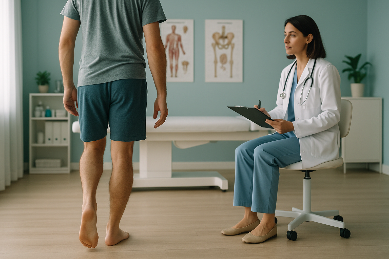
A Trendelenburg gait looks like a dip or drop of the pelvis on the side of the swinging leg. When you stand on one foot, the hip abductors on the standing side should keep the pelvis level. If those muscles are too weak or inhibited, the unsupported side drops. The body often leans sideways to make up for it. When this happens with every step, you get a clear side-to-side sway, sometimes called a waddling gait.
The Trendelenburg sign is the clinical test behind the name. A clinician asks you to stand on one leg and watches whether the opposite hip drops. A positive result does not tell you why the muscles are failing. It only confirms that they are.
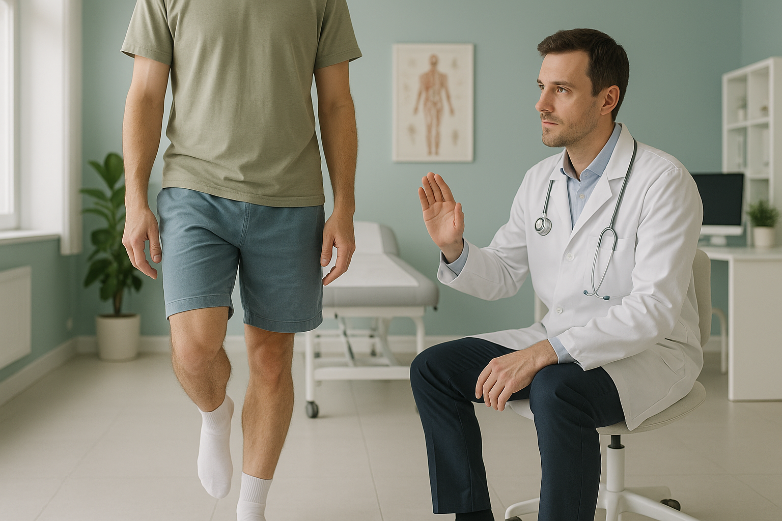
There is also something called a compensated Trendelenburg gait. Instead of the pelvis dropping, you lean your upper body over the weak hip to keep your balance. Over time, this shift of the trunk can put extra stress on your spine and the opposite hip.
How do you know if your hip drops when you walk?
You may not realise your pelvis is dropping. Many people just feel that something is "off" about their walking, but cannot say exactly what. These are the common signs.
Other people may notice a side-to-side sway before you feel it yourself.
Clothes that sit lower on one side can hint at a lateral pelvic tilt.
The hip doing the extra work often becomes sore at the outer buttock or greater trochanter.
The spine may curve to make up for the pelvic drop. This can cause pain away from the hip itself.
If you also notice numbness, weakness in the leg, or any loss of bladder or bowel control, get urgent medical help from your doctor or the nearest emergency department rather than booking a scan.
What causes a Trendelenburg gait?
A Trendelenburg gait happens when the hip abductors, mainly the gluteus medius, cannot keep the pelvis stable while you stand on one leg. Several things can lead to this.
Long hours of sitting, inactivity or not using the muscles after surgery can weaken them significantly.
Conditions such as hip dysplasia in adults or advanced wear of the joint can change how the muscles around the joint work.
Damage to or compression of the superior gluteal nerve stops the signal reaching the hip abductors.
Even a small difference in leg length can tilt the pelvis and overload one side.
Surgery may change the muscle balance around the hip, for a while or permanently.
The body works as a connected chain from feet to head. So a weakness at the hip can force the knee, spine or opposite hip to make up for it. Pain often shows up away from the real source.
Why does imaging done lying down miss a Trendelenburg pattern?
Standard X-rays, CT and MRI scans are taken while you lie flat. In that position, gravity is taken away and your muscles relax. The pelvic drop that defines a Trendelenburg gait disappears, because you are not bearing weight. A scan may show the hip joint looks "normal", even though your walking pattern is clearly abnormal.
An EOS scan captures the pelvis, hips and full legs while you stand. Your muscles are working and gravity is acting on your skeleton. Any pelvic tilt, leg length difference or uneven joint is visible in its true, loaded state. This is how ScanAlign standing EOS imaging shows alignment problems that other scans can miss.
Imaging done lying down is excellent for seeing bone detail and soft tissue. But it removes the very force that causes the pelvis to drop: your body weight. A hip joint that looks well aligned on a supine X-ray may sit very differently once you stand up and walk.
When MRI is the better choice: EOS shows bones, joints and alignment under load. If your clinician suspects a soft-tissue injury, a torn labrum, a disc problem or nerve compression, MRI is usually the right tool for that job.
A full standing image also supports gait analysis. It gives clinicians a structural baseline to compare with what they see when you walk.
What can be done about a Trendelenburg gait?
The right approach depends entirely on the cause behind it. ScanAlign provides the diagnostic imaging that helps find that cause. It does not provide treatment. These are the general directions a clinician may take once the cause is clear.
- Targeted strengthening. If the gluteus medius is weak but the joint and nerve are intact, a structured physiotherapy programme focused on the hip abductors is often the starting point.
- Dealing with a leg length difference. Where imaging confirms a meaningful difference, an orthotist or surgeon can discuss options, from a simple heel lift to surgical correction.
- Care focused on the joint. If the hip joint itself is the problem, an orthopaedic assessment may be needed to decide whether conservative management or surgery is right.
- Rehabilitation after surgery. After a hip replacement, specific rehab protocols target the hip abductors to restore an even walking pattern.
In every case, accurate imaging of the whole chain, pelvis, hips and legs, while you bear weight, gives clinicians the clearest picture of what needs attention first.
So before you start treatment, or change the one you are on, it is worth getting a detailed diagnosis. Knowing whether the cause sits in the pelvis, the legs or the spine changes what will actually help you.
Wondering whether your hip drop is structural? A standing EOS scan can show you.
Book a free video consultationFrequently asked questions about Trendelenburg gait
It is a walking pattern where one side of your pelvis drops when you step onto the other leg. It means the muscles of the standing hip are not strong enough to keep the pelvis level.
The Trendelenburg sign is a clinical test. You stand on one leg and a clinician watches whether the opposite hip drops. A positive sign confirms hip abductor weakness, but it does not explain the cause.
The most common reason is weakness of the gluteus medius muscle. Other causes include hip joint problems, nerve damage, differences in leg length and changes after hip surgery.
They overlap, but they are not the same. A waddling gait can have several causes. When the waddle is caused specifically by hip abductor weakness and pelvic drop, it fits the Trendelenburg pattern.
A standard X-ray taken lying down removes gravity and muscle load. So it cannot show the pelvic drop that defines this walking pattern. A standing, weight-bearing scan shows far more about alignment problems.
An EOS scan captures your skeleton from head to toe while you stand, using about 90% less radiation than a standard X-ray, around ten times less. It shows how your joints, pelvis and legs line up under your own body weight.
No. ScanAlign is a private self-pay clinic. You can refer yourself directly and start with a free video consultation to discuss whether a scan is right for you.
Yes. EOS uses about 90% less radiation than a standard X-ray, around ten times less. This makes it one of the lowest-dose options available for full skeletal imaging.
That depends on the cause. Many cases improve significantly with targeted physiotherapy. Others may need further treatment. The first step is always an accurate diagnosis through proper imaging and clinical assessment.
The price for patients travelling from abroad is confirmed during your free video consultation, along with exactly what is included. Our frequently asked questions page answers other common questions.
See what standing imaging shows about your hip
A free video consultation is the easiest way to find out whether a standing EOS scan in London is right for you, before you plan your trip. No referral needed, no obligation.
Book a free video consultationWritten by Abbas Dhami (Specialist Diagnostic Radiographer)
Sources
- Hardcastle P, Nade S. The significance of the Trendelenburg test. Journal of Bone and Joint Surgery (British Volume). 1985;67-B(5):741-746.
- NHS. Hip pain in adults. nhs.uk. Available at: https://www.nhs.uk/conditions/hip-pain/
- Illés T, Somoskeöy S. The EOS imaging system and its uses in daily orthopaedic practice. International Orthopaedics. 2012;36(7):1405-1411.
This page is general information and not a diagnosis.
ScanAlign is the trading name of CoreMed Solutions Ltd and operates under The Harley Street Hospital's CQC licence.