
How Does Standing Imaging Support Osteopathic Assessment?
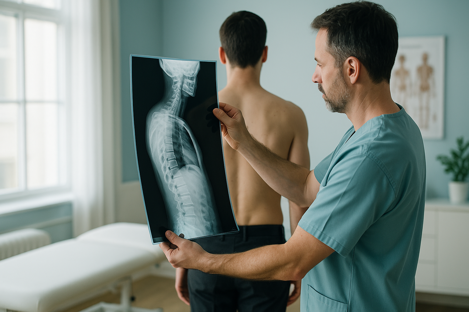
Standing whole-body imaging gives osteopaths an objective, weight-bearing view of alignment from head to feet. It shows how gravity loads the skeleton in normal posture. It reveals compensation patterns, leg-length differences and pelvic obliquity that lying-down scans can miss. The result is a measurable baseline that supports and refines your clinical findings.
Key points
- Standing EOS imaging shows the skeleton under real weight-bearing load, not relaxed on a table.
- It captures the full kinetic chain in a single exposure, with about 90% less radiation than a standard X-ray, around ten times less.
- Osteopaths can refer patients directly through a simple registration process.
- Reports include measurements that add to your hands-on assessment and help you explain findings to patients.
- EOS shows bones, joints and alignment. For soft tissue, discs or nerves, MRI is usually the better tool.
Why does standing imaging matter for osteopathic assessment?
Standing imaging matters because your patients are not lying down when they have their symptoms. Most conventional imaging, whether X-ray, CT or MRI, is taken with the patient supine. In that position gravity is removed and muscles relax. The spine, pelvis and lower limbs settle into a shape that may look quite different from the one causing pain.
As an osteopath, you assess patients standing, walking and bending. You feel asymmetries, notice compensations and build a clinical picture based on function under load. Standing imaging does the same thing radiologically. It captures the skeleton in its habitual posture, so the image matches what your hands tell you.
What does a whole-body standing scan show an osteopath?
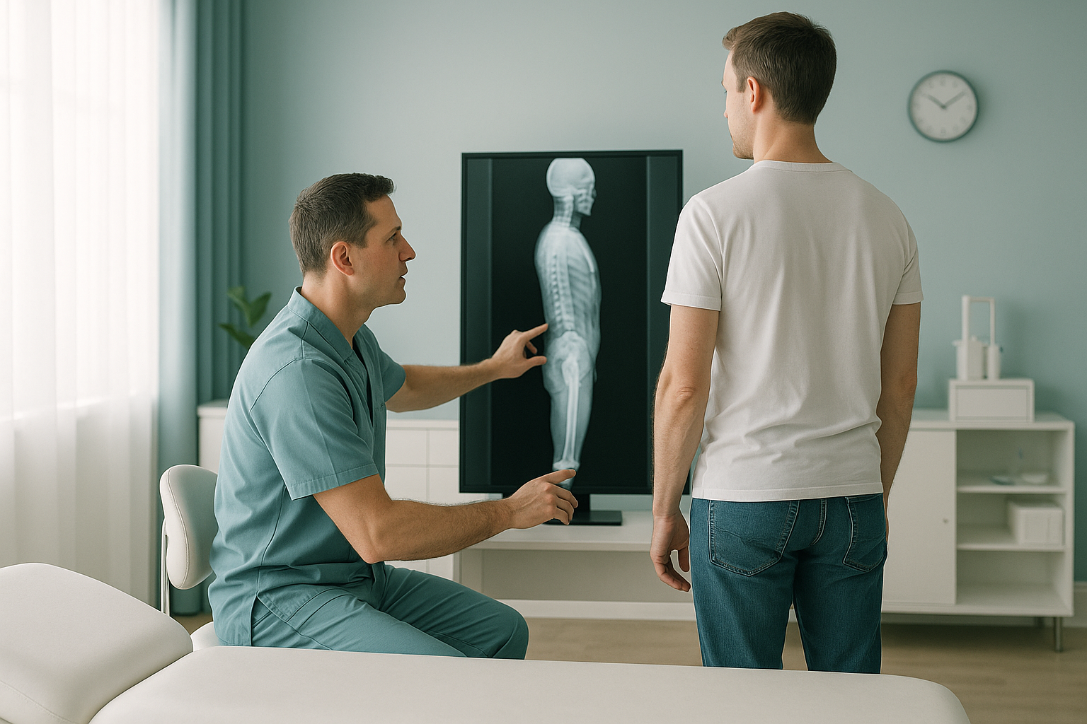
A whole-body standing scan shows the entire skeleton, from the skull base to the feet, in a single calibrated image. You can see how one region affects another. This is the whole-person approach at the heart of osteopathic thinking.
Because the image captures the full kinetic chain, you can trace a compensation from where it starts to where the patient feels pain. A pelvic tilt, for example, may be the body's response to a difference further down the chain. A single-region X-ray would never reveal this.
Can osteopaths refer patients for imaging in the UK?
Yes. In the UK, osteopaths registered with the GOsC can refer patients for imaging. At ScanAlign, whole-body standing imaging, the referral process is simple and designed for allied health professionals.
Fill in a short form to register to refer patients. We verify your GOsC registration and set up your account.
Send a referral with the clinical question you want answered. No letter from the patient's doctor is needed.
The scan takes place standing up. It is quick, comfortable and uses about 90% less radiation than a standard X-ray, around ten times less.
You receive a detailed radiology report with measurements that map directly onto your clinical findings.
Want to discuss a case before referring? We offer a free video consultation so you can check whether imaging is appropriate.
Book a free video consultationWhat is the difference between standing and lying-down imaging?
The key difference is gravity. When your patient lies down, muscles relax, the pelvis levels out and spinal curves flatten. The image may look reassuringly normal, even when the patient has significant pain under upright load.
Weight-bearing and functional. The skeleton is loaded exactly as it is in daily life. Pelvic obliquity, scoliotic curves and leg-length differences are captured as your patient really lives with them. The full body is imaged in one exposure, so you can trace compensation patterns across regions.
Relaxed and unloaded. Conventional X-rays and most MRI or CT scans are taken lying down. Gravity is removed, so the body settles into a neutral shape. Asymmetries can reduce or disappear. Single-region images also make it harder to see how one area is affecting another.
Standing imaging does not replace lying-down scans. It adds a functional view that is especially relevant to the way osteopaths assess and reason.
Which patients benefit most from a standing scan referral?
Any patient where you suspect alignment or structure is contributing to their symptoms may benefit. Here are some common situations.
- Persistent low back or pelvic pain that does not respond as expected
- Suspected leg-length difference or pelvic obliquity
- Recurring problems that suggest a compensatory pattern
- Assessment of hip or knee alignment before or after surgery
- Patients who want an objective baseline before a course of treatment
- Suspected disc herniation, nerve root compression or spinal cord pathology
- Soft-tissue injuries (muscle, tendon, ligament)
- Acute trauma needing urgent assessment
- Red-flag symptoms such as sudden weakness, numbness, or loss of bladder or bowel control
A note on MRI. EOS imaging shows bones, joints and alignment clearly. For soft tissue, discs or nerves, MRI is usually the better tool. Many patients benefit from both, depending on the clinical question.
Red-flag reminder. If your patient has sudden weakness, numbness, or loss of bladder or bowel control, please tell them to seek urgent medical care from their doctor or their local emergency services, rather than booking a scan.
Before a patient starts treatment, or changes the one they are on, it is worth getting a detailed diagnosis. Knowing whether the cause sits in the pelvis, the legs or the spine changes what will actually help.
Frequently asked questions
Yes. In the UK, GOsC-registered osteopaths can refer patients for imaging. At ScanAlign you can refer directly, without a letter from the patient's doctor, once you have completed a short registration.
Visit the registration page and fill in a short form. We verify your professional registration and set up your account, usually within a working day.
EOS is a low-dose, standing X-ray system that captures the full skeleton from head to feet in a single exposure. It uses about 90% less radiation than a standard X-ray, around ten times less.
The radiation dose is very low, about 90% less than a standard X-ray. The scan is non-invasive and takes only a short time. Patients stand comfortably throughout.
EOS is designed for bones, joints and alignment. It does not image soft tissue in detail. If you suspect a disc, ligament or nerve problem, MRI is usually the better choice.
ScanAlign is a self-pay clinic, and the price for patients travelling from abroad is confirmed during the free video consultation. You can refer a patient here.
The radiology report includes relevant alignment measurements across the spine, pelvis and lower limbs. These are calibrated to the standing position and can be tracked over time.
Absolutely. A full-body standing image is one of the clearest ways to help a patient see their alignment and understand why you are recommending a particular approach.
At 19 Harley Street, London, within The Harley Street Hospital. The clinic is easy to reach, and the setting is comfortable and professional.
Yes. You can book a free video consultation to discuss whether imaging is appropriate for a particular patient before you make a referral.
Ready to refer, or want to discuss a case?
Book a free video consultation to talk through whether standing imaging would add value for your patient.
Book a free video consultationSources
- Dubousset, J. et al. "EOS: a new imaging concept." European Spine Journal, 2010. Overview of EOS low-dose biplanar imaging technology and clinical validation.
- General Osteopathic Council (GOsC). Osteopathic Practice Standards, 2023. Sets out the scope of practice and referral rights for UK-registered osteopaths.
- NHS England. Clinical Imaging: Ionising Radiation Dose Comparisons. Public guidance on typical radiation doses from common imaging procedures.
Written by Abbas Dhami (Specialist Diagnostic Radiographer)
This page is general information and not a diagnosis.
ScanAlign is the trading name of CoreMed Solutions Ltd and operates under The Harley Street Hospital's CQC licence.