
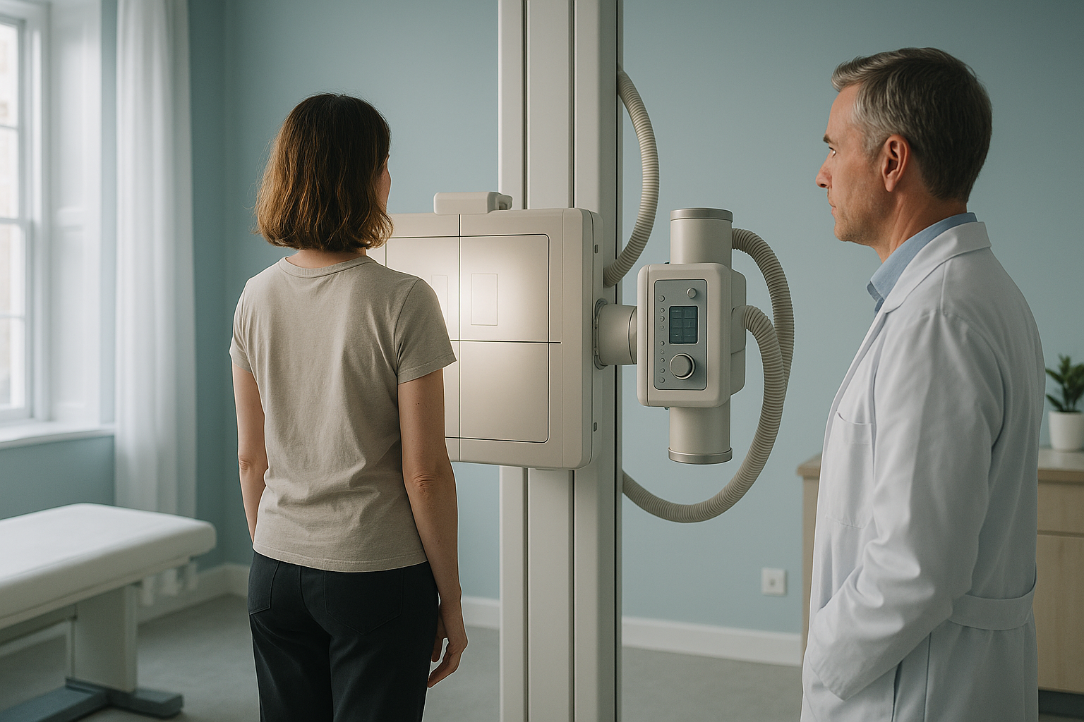
When Does Standing Imaging Help in General Practice?
Standing whole-body EOS imaging is most useful when a patient's bone, joint or muscle symptoms continue after conservative management, and you suspect an alignment or biomechanical cause that lying-down (supine) imaging cannot show. It does not replace MRI for soft-tissue assessment. It is not indicated for acute presentations or red-flag symptoms.
Key points
- Standing imaging shows weight-bearing alignment that lying-down scans can miss.
- It is best suited to persistent musculoskeletal pain after a trial of conservative management, not to acute or red-flag presentations.
- EOS delivers about 90% less radiation than a standard X-ray, around ten times less.
- Reports are sent directly to you, the referring doctor, to support onward specialist referral or management.
- Patients can refer themselves, but a referral from their doctor sharpens the clinical question and makes the report more useful.
This page is written for GPs, family doctors and other primary care clinicians. It is deliberately cautious. Not every patient with back or joint pain needs imaging. We would rather explain when standing imaging is not indicated than oversell when it is.
When should a doctor consider referring for standing imaging?
Standing whole-body imaging is worth considering when symptoms continue after a proper trial of conservative management (typically six weeks or more). It fits when you suspect the clinical picture involves alignment, a leg-length difference or biomechanical compensation, rather than a problem in the soft tissue alone.
In each of these cases, the clinical question is about how the skeleton behaves under load, not about soft tissue. That is where standing imaging is strongest.
When is standing imaging not the right choice?
Standing EOS imaging is not right for every musculoskeletal complaint. It is important to be clear about its limits. That way, neither you nor your patient spends time or money on a test that will not answer the clinical question.
- Acute injury, fracture or red-flag symptoms (cauda equina, progressive neurological deficit, unexplained weight loss, fever)
- Suspected disc herniation, nerve root compression or spinal cord pathology (MRI is the right test)
- Soft-tissue injuries: ligament tears, meniscal damage, tendinopathy
- Non-specific low back pain within the first six weeks, where NICE guidance in the UK recommends conservative management before imaging
- Patients who cannot stand without support for the length of the scan
- Chronic back, hip or knee pain that continues after conservative management
- Clinical suspicion of an alignment-related cause (pelvic tilt, leg-length difference, adult scoliosis)
- Pre-operative alignment assessment requested by a specialist
- Post-operative review of joint replacement or spinal instrumentation under weight-bearing conditions
- Musculoskeletal symptoms in several places that suggest a compensation chain
A note on MRI. EOS imaging shows bones, joints and alignment under load. For soft tissue, discs or nerves, MRI is usually the better tool. In many cases, the two work together rather than compete. If your clinical question is about disc pathology or nerve compression, MRI is still the first choice.
Red-flag reminder. If your patient has sudden weakness, loss of bladder or bowel control, saddle anaesthesia or rapidly progressive neurological deficit, they need urgent medical care through the nearest emergency department, not imaging at a private clinic.
What does standing imaging show that lying-down scans miss?
When a patient lies down for a conventional X-ray, CT or MRI, gravity is taken away. Muscles relax. The spine, pelvis and legs settle into a position that may look normal. But it does not show how the skeleton really behaves when standing and walking.
The patient stands naturally inside the EOS system. The scan captures the whole skeleton from head to feet in a single pass. Because the patient is bearing weight, the image shows real-world alignment: how the spine curves, how the pelvis sits, how the knees and ankles share the load. A pelvic tilt or leg-length difference that disappears on the couch becomes visible. The body is a connected chain, and compensation patterns from feet to head are captured in one image.
EOS uses about 90% less radiation than a standard X-ray, around ten times less, which matters when full-body imaging is involved.
Lying-down imaging is excellent for soft tissue (MRI) and for acute bone injury (CT or conventional X-ray). But it cannot show alignment under load. A pelvis that tilts when standing may look level on the couch. A compensatory spinal curve may flatten out when gravity is taken away. If the clinical question is about alignment or biomechanics, a supine image may look normal even when a standing image would not.
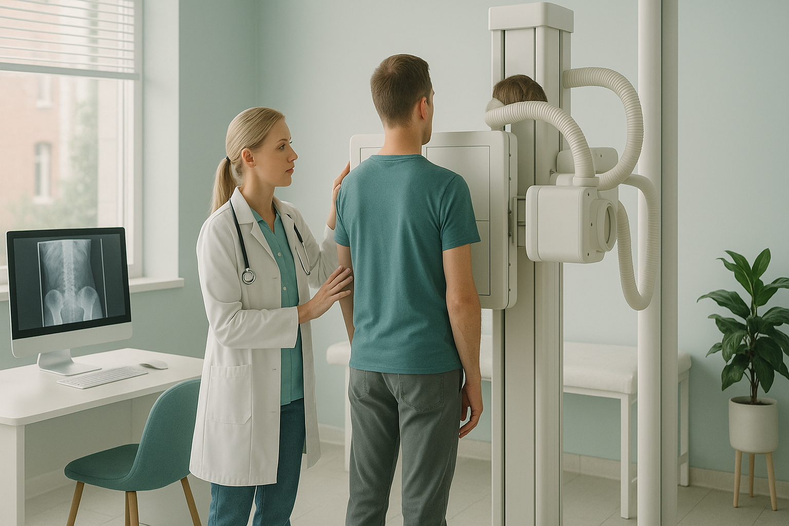
This does not mean lying-down imaging is wrong. It answers a different question. When you need to know about alignment and load, standing imaging fills the gap.
How does the referral process work?
Referring a patient is simple. No special forms, no health-system approvals and no waiting for funding approval. Patients pay privately for the scan.
Fill in a short online form to register to refer patients. You only need to do this once.
Send a referral with the clinical question and relevant history. You can refer a patient online or by email. A clear clinical question helps us write a more focused report.
The patient comes to our clinic at 19 Harley Street, London, for a standing EOS scan. It takes only a few minutes. No contrast, no preparation, no sedation.
A full report is sent to you as the referring doctor, with measurements and findings relevant to your clinical question. You can then use it to support onward specialist referral or adjust management.
Patients can also refer themselves. Many do. But a referral from their doctor adds value because it frames the clinical question, so the report covers exactly what you need to know.
What does the doctor receive in the imaging report?
You receive a structured radiology report with standing alignment measurements of the spine, pelvis and legs. The report is shaped around the clinical question you give at referral.
If the report finds something outside the scope of what we image (for example, if soft-tissue assessment is needed), we will say so clearly. The report is built to be practical: something you can use in a follow-up consultation, attach to a specialist referral letter or discuss with the patient directly.
Before a patient starts treatment, or changes the one they are on, a detailed diagnosis is worth having. Knowing whether the cause sits in the pelvis, the legs or the spine changes what will actually help.
For more on what patients go through during the scan, see our back pain clinic page.
If you have a patient who might benefit from standing alignment imaging and would like to talk through the case informally first, you are welcome to get in touch.
Refer a patientFrequently asked questions from doctors
No. Patients can refer themselves. But a referral from their doctor improves the process. It gives us a clinical question to focus the report on, which makes the findings more useful to you and to any specialist involved later.
Yes. EOS delivers about 90% less radiation than a standard X-ray, around ten times less. This matters most for full-body imaging, where conventional X-rays would need several exposures at higher doses.
The price for patients travelling from abroad is confirmed during the free video consultation.
Yes. The report is a formal radiology document that can be attached to a referral letter. Many doctors use it to strengthen a case for orthopaedic or spinal specialist review.
A standard X-ray is usually taken lying down and covers one area at a time. EOS captures the whole skeleton from head to feet in a single standing image, at a much lower radiation dose. This gives a complete picture of alignment under load, which imaging one section at a time cannot give.
If your clinical question is about soft tissue, discs, nerves or spinal cord pathology, MRI is the right choice. EOS is for skeletal alignment while bearing weight. In some cases, both are useful and work together.
NICE in the UK recommends against routine imaging for non-specific low back pain in the first six weeks. After that, if symptoms continue and a structural or alignment cause is suspected, imaging becomes appropriate. Standing EOS fits into this pathway as a tool for looking into biomechanical questions that conservative management has not resolved.
You can register online in a few minutes. Once registered, you can refer patients directly. There is no cost or obligation to register.
You are welcome to contact us to discuss a case before referring. We would rather help you choose the right type of imaging than accept a referral that will not answer your question.
We are at 19 Harley Street, London, operating under The Harley Street Hospital's CQC licence. No special preparation is needed. The patient should wear comfortable clothing and be able to stand still for a few minutes.
Ready to refer a patient?
Register once, then refer patients online. Reports are sent directly to you. No cost to register.
Register to refer patients Refer a patientAbout ScanAlign
ScanAlign, standing EOS imaging in London, is a private self-pay imaging clinic at 19 Harley Street. We provide standing whole-body EOS imaging to help clinicians and patients understand musculoskeletal alignment under real-world, weight-bearing conditions. We are a diagnostic imaging service only and do not provide treatment.
Sources
- NICE guideline NG59: Low back pain and sciatica in over 16s: assessment and management. National Institute for Health and Care Excellence, 2016 (updated 2020).
- Deschênes S, et al. Diagnostic imaging for the assessment of a scoliosis: reduction of radiation dose. Spine, 2010; 35(9): 989-994.
- Royal College of Radiologists. iRefer: Making the best use of clinical radiology. 8th edition, 2017.
Written by Abbas Dhami (Specialist Diagnostic Radiographer).
This page is general information and not a diagnosis.
ScanAlign is the trading name of CoreMed Solutions Ltd and operates under The Harley Street Hospital's CQC licence.