
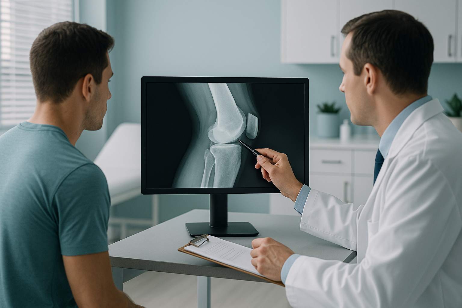
How Does Imaging Support Sports and Exercise Medicine?
Sports medicine imaging gives consultants a whole-body view of bone alignment, joint loading and asymmetry while the body carries its own weight. Standing EOS imaging captures the full kinetic chain in one low-dose scan. It shows compensatory patterns that lying-down scans can miss. This helps guide return to play decisions, the investigation of repeated injuries and baseline screening for a squad.
Key points
- Standing whole-body imaging shows how bones and joints really take load under body weight.
- The kinetic chain from the feet to the spine is captured in one scan. This shows asymmetry and compensatory tilt.
- EOS uses about 90% less radiation than a standard X-ray, around ten times less.
- The results add to MRI, which is still the better tool for checking soft tissue, discs and nerves.
- No referral from a family doctor is needed. SEM consultants can refer athletes directly.
If you are a sports and exercise medicine (SEM) consultant, you already know that many bone, joint and muscle complaints in athletes are not isolated injuries. They come from load, and from how the whole body lines up and moves under stress. Standard imaging often looks at one area on its own, and usually while the patient is lying down. This can miss the very alignment problems that explain why an injury keeps coming back.
At ScanAlign, standing EOS imaging in London, we take full-length images of the skeleton from head to feet in one capture, while the body carries its own weight. This gives SEM consultants the biomechanical context they need, alongside the usual MRI and ultrasound findings.
Why does standing imaging matter for athletes?
Standing imaging matters because gravity and working muscles change the way joints line up. When an athlete lies down for a standard X-ray, CT or MRI, the muscles relax. The spine, pelvis and legs settle into a position that does not reflect real-life loading. A mild difference in leg length, a rotational tilt of the pelvis or an uneven valgus at the knee may flatten out on the table. It can then come back the moment the athlete stands and trains.
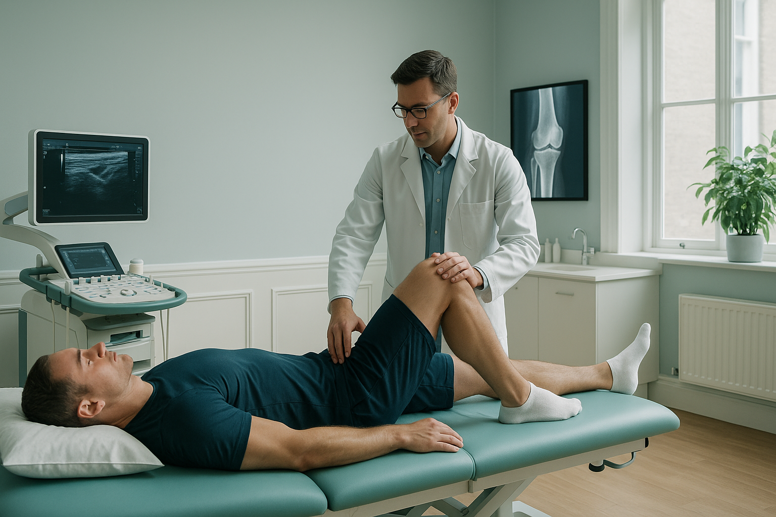
EOS imaging solves this by scanning the athlete upright, in a natural standing position. The system captures the whole skeleton in one go. So the link between the spine, pelvis, hips, knees and ankles can be seen in a single image. Rotational profiles, differences in limb length and frontal-plane asymmetry can all be measured, and the measurements can be repeated.
What clinical questions can standing alignment imaging answer?
Standing alignment imaging answers questions about how the whole kinetic chain spreads load, not just what one joint looks like on its own. These are the cases SEM consultants most often use it for.
A hamstring or calf tear that keeps coming back may be driven by pelvic obliquity or uneven limb length. Whole-body alignment data shows the biomechanical context behind the repeated pattern.
After a significant leg or spinal injury, standing imaging gives objective alignment data. This helps the consultant judge whether the athlete's structure is ready for full training loads.
Groin, hip or knee pain that gets worse as training volume rises may relate to frontal-plane malalignment or rotational asymmetry. These patterns only become visible when the skeleton is under load.
A standing scan before the season creates a baseline for comparison. If an injury happens during the season, you have a reference point to measure what has changed and by how much.
To read more about how alignment data helps with prevention, see our guide to sports injury prevention.
How does standing imaging compare with standard scans?
Standing EOS imaging and standard imaging do different jobs. Knowing what each one offers helps you pick the right tool for each clinical question.
Full kinetic chain in one image. The spine, pelvis, hips, knees and ankles are captured at the same time, while the athlete stands under body weight.
Low radiation dose. About 90% less radiation than a standard X-ray, around ten times less. Suitable for baseline and follow-up scans.
Repeatable measurements. The standing position is standardised, so scans taken months apart can be compared reliably.
Focus on one area. A standard X-ray, CT or MRI usually images one area. This is useful for diagnosing a specific fracture or soft-tissue injury. But it does not show how that area relates to the rest of the skeleton.
No body-weight load. The body relaxes on the table. Alignment differences caused by gravity, muscle tension and loading do not show up.
Essential for soft tissue. MRI is the gold standard for checking tendons, ligaments, cartilage, discs and nerves. EOS does not replace that.
EOS and MRI work together, not against each other. EOS shows bones, joints and alignment under load. For soft tissue, discs or nerves, MRI is usually the better tool. Many SEM consultants ask for both: MRI to check the local tissue injury, and EOS to understand the biomechanical setting around it.
How do SEM consultants refer an athlete?
The referral process is simple. No letter from a family doctor is needed. SEM consultants, team doctors and club medical staff can refer athletes directly.
Create your referrer profile once. This lets you register to refer patients and manage referrals easily.
Send us the athlete's details and your clinical question. You can refer an athlete online at any time.
The athlete comes to our London clinic for a standing EOS scan. It takes only a few minutes and uses about 90% less radiation than a standard X-ray.
A detailed alignment report is sent directly to you. It covers the measurements and findings that relate to your clinical question.
Which athletes benefit most from standing alignment imaging?
The athletes who benefit most have long-lasting, load-related or repeated bone, joint or muscle problems, where imaging of one area has not given a full explanation.
- Repeated hamstring, groin or calf injuries
- Long-lasting hip or knee pain made worse by training load
- Athletes after surgery who are nearing return to play
- Squad screening before the season, for baseline data
- Scoliosis or spinal asymmetry in adult athletes
- You need detail of soft tissue, discs or nerves (MRI is better)
- There is a suspected acute fracture that needs urgent assessment
- The athlete has numbness, weakness, or loss of bladder or bowel control. These are red flags that need urgent medical attention (contact a doctor or go to the nearest emergency department)
Before an athlete starts treatment, or changes the one they are on, it is worth getting a detailed diagnosis. Knowing whether the cause sits in the pelvis, the legs or the spine changes what will actually help.
Frequently asked questions
Sports medicine imaging is used to check how an athlete's skeleton lines up under load. It helps SEM consultants understand repeated injuries, load-related pain, asymmetry and readiness to return to play. It does this by showing the full kinetic chain in one standing image.
An EOS scan images the whole body while you stand. It captures everything from head to feet in one pass. It uses about 90% less radiation than a standard X-ray, around ten times less. A normal X-ray usually focuses on one area and may be taken lying down.
No. SEM consultants, team doctors and club medical staff can refer athletes directly. There is no need for a letter from a family doctor.
Yes. EOS uses about 90% less radiation than a standard X-ray, around ten times less. So it is well suited to baseline and follow-up scans within the same season.
No. EOS shows bones, joints and alignment. For soft tissue such as ligaments, tendons, cartilage, discs or nerves, MRI is usually the better tool. Many consultants use both together for a fuller picture.
The scan itself takes only a few minutes. The athlete stands still inside the EOS system while the image is taken. No special preparation is needed.
The report includes whole-body alignment measurements for the spine, pelvis, hips, knees and legs. It points out asymmetry, compensatory patterns and any clinically significant findings. All of these relate to the clinical question you send with the referral.
Yes. A standing scan before the season gives a baseline. If an injury happens during the season, the baseline lets you measure what has changed. This makes it easier to guide rehabilitation and return to play.
The price for patients travelling from abroad is confirmed during the free video consultation. Squad screening can also be discussed then.
ScanAlign is at 19 Harley Street, London, within The Harley Street Hospital. It is a private, self-pay clinic. SEM consultants who wish to refer athletes do not need a family doctor's referral.
Ready to refer an athlete?
Get the whole-body alignment data you need to support your clinical decisions. No family doctor's referral needed.
Book a free video consultationSources
- Illés, T. & Somoskeöy, S. (2012). The EOS imaging system and its uses in daily orthopaedic practice. International Orthopaedics, 36(7), 1405–1411.
- Brink, M.S. et al. (2010). Monitoring load, recovery, and performance in young elite soccer players. Journal of Strength and Conditioning Research, 24(3), 597–603.
- British Association of Sport and Exercise Medicine (BASEM). Position statements on musculoskeletal assessment in sport. basem.co.uk.
Written by Abbas Dhami (Specialist Diagnostic Radiographer).
This page is general information and not a diagnosis.
ScanAlign is the trading name of CoreMed Solutions Ltd and operates under The Harley Street Hospital's CQC licence.