
Perthes Disease: What Is It and Why Does Your Child Need Repeat Imaging?
Perthes disease (also called Legg-Calvé-Perthes disease) is a hip condition of childhood. The blood supply to the ball of the thigh bone is cut off for a while. The bone softens, changes shape and then slowly rebuilds over several years. Because recovery is slow, children need regular scans to follow the healing. This is why low dose scanning is important.
Key takeaways
- Perthes disease most often affects children between the ages of 4 and 10.
- The femoral head (ball of the hip) loses its blood supply for a while and then regrows.
- Recovery takes two to four years, with regular scans needed at every stage.
- Radiation from repeat X-rays adds up and is a real concern. Low dose EOS imaging uses about 90% less radiation than a standard X-ray.
- Diagnosis and treatment decisions are made by a paediatric orthopaedic team, not an imaging clinic.
What are the symptoms of Perthes disease?
The first sign is usually a limp that comes and goes. Sometimes there is a mild ache in the hip, groin or knee. The pain can show up in the knee rather than the hip, so Perthes disease is sometimes missed at first.
If your child has a limp that does not clear up within a week or two, or complains of hip or knee pain with no clear injury, see your child's doctor. They can arrange the first check-up.
What causes Perthes disease?
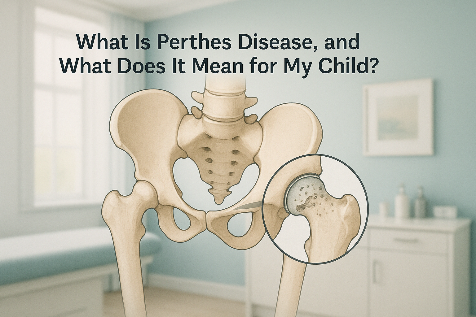
The cause is a short-term break in the blood supply to the femoral head, the rounded top of the thigh bone that sits inside the hip socket. Without enough blood, the bone weakens (a process sometimes called avascular necrosis). Nobody knows exactly why this happens. It is not caused by injury, diet or anything a parent did or did not do.
What are the stages of Perthes disease?
Perthes disease passes through four known stages over roughly two to four years. Each stage looks different on scans. This is why regular scans are needed to follow progress and guide the orthopaedic team's decisions.
Initial (necrosis)
The blood supply is cut off. The femoral head starts to soften, though its outer shape may still look normal on early X-rays.
Fragmentation
The body starts to break down and absorb the weakened bone. On scans the femoral head can look patchy or broken up. This is actually the start of healing.
Re-ossification
New, healthy bone slowly replaces the old. The shape of the femoral head as it regrows is what decides the long-term outlook.
Healed (residual)
Bone growth is complete. The final shape of the femoral head, round or flattened, affects how the hip works into adult life.
How is Perthes disease monitored with imaging?
Serial imaging, meaning repeat scans taken at set times over months and years, is the backbone of Perthes care. Scans show the orthopaedic team how the femoral head is reshaping, whether it is staying well seated in the socket, and whether the treatment plan needs to change.
A child with Perthes disease may need many sets of hip images over the full course of the condition. That is a lot of radiation if each session uses a standard X-ray dose. This is where the total, built-up dose matters most.
Reducing the total dose matters. EOS imaging uses about 90% less radiation than a standard X-ray, around ten times less. Over two to four years of monitoring, that difference adds up a great deal. Learn more about EOS scan safety and radiation.
Your child's paediatric orthopaedic consultant decides when each scan is needed and which views are required. Paediatric imaging at ScanAlign simply provides the images at a much lower radiation dose, then sends the results to your child's medical team.
Why does low dose imaging matter for children with Perthes disease?
Children are more sensitive to radiation than adults, because their cells are still dividing quickly as they grow. The more scans a child has, the more the doses add up. Perthes disease needs years of follow-up, so keeping each single dose as low as possible is a sensible precaution.
EOS captures the hip and pelvis in a single standing sweep using about 90% less radiation than a standard X-ray, around ten times less. Over many appointments, that reduction becomes meaningful. Standing imaging also shows how the hip behaves under the child's own body weight. This can be useful for judging how the joint works in daily life.
ScanAlign, low dose standing imaging, is available at 19 Harley Street, London. You can book a paediatric monitoring scan once your child's consultant has asked for follow-up imaging.
A note on MRI. EOS shows bones, joints and alignment clearly. For soft-tissue detail, such as blood flow within the femoral head or changes in the cartilage, MRI is usually the better tool. Your child's orthopaedic team will choose the right type of scan for each stage. The two often work alongside each other.
What is the long-term outlook for Perthes disease?
The outlook depends mainly on two things: the age at which the condition starts, and how round the femoral head is when it finishes regrowing. Younger children (under six at onset) tend to do better. They have more growing time for the bone to reshape into a good form.
Many children recover with a hip that works well and causes little or no trouble into adult life. Some are left with a femoral head that is flatter or less even. This can affect the range of movement and may raise the chance of hip problems later in life.
What helps during recovery?
The paediatric orthopaedic team may advise physiotherapy to keep the hip moving, periods of rest or less activity, or in some cases a brace or surgery to keep the femoral head well held in the socket. These are called containment treatments. ScanAlign does not provide treatment, but the images we produce support the team making those decisions.
Before your child starts a treatment, or changes the one they are on, it is worth getting a detailed diagnosis. Knowing whether the cause sits in the pelvis, the legs or the spine changes what will actually help.
Frequently asked questions about Perthes disease
It most often appears between the ages of 4 and 10, with a peak around age 5 to 7. It is more common in boys than girls.
Yes. The full medical name is Legg-Calvé-Perthes disease, named after the three doctors who described it. Most people shorten it to Perthes disease.
In about 10 to 15 per cent of cases, both hips are involved, usually at different times rather than at the same time.
It varies, but monitoring usually runs for two to four years, with scans at regular intervals. That can mean quite a few sessions in total. This is why keeping each dose low really matters.
EOS uses a narrow slot-scanning beam rather than a broad burst of radiation. This delivers about 90% less radiation than a standard X-ray, around ten times less per session.
No formal referral is needed. However, Perthes disease is managed by a paediatric orthopaedic team. So we recommend working with your child's consultant, so the right views are requested and the results go straight to them.
The scan is very quick. Most children can stand comfortably for the few seconds it takes. If your child has difficulty putting weight on the leg, tell the team when you book and they will advise on the best approach.
They do different things. EOS is excellent at showing bone shape and joint alignment. MRI shows soft tissue detail, including blood flow within the femoral head. Your child's team may use both at different stages.
The price for patients travelling from abroad is confirmed during your free video consultation.
If the femoral head heals in a round shape, the risk of early arthritis is low. A less round shape can raise the risk of hip problems in adult life, but many people live active lives regardless. Good monitoring during childhood helps the orthopaedic team make decisions that improve the long-term outcome.
Years of scans. A fraction of the dose.
If your child is being monitored for Perthes disease, a free video consultation can help you understand whether low dose EOS imaging suits their follow-up plan.
Book a free video consultationSources
- NHS. "Perthes' disease." nhs.uk, reviewed 2022.
- Joseph B. "Natural history of early onset and late-onset Legg-Calve-Perthes disease." Journal of Pediatric Orthopaedics, 2011; 31(2 Suppl): S152-S155.
- Deschênes S et al. "Diagnostic imaging of the pediatric hip: EOS, radiography, and MRI." Pediatric Radiology, 2013; 43(Suppl 1): S68-S73.
Written by Abbas Dhami (Specialist Diagnostic Radiographer)
This page is general information and not a diagnosis.
ScanAlign is the trading name of CoreMed Solutions Ltd and operates under The Harley Street Hospital's CQC licence.