
What Is a Periacetabular Osteotomy (PAO) for Hip Dysplasia?
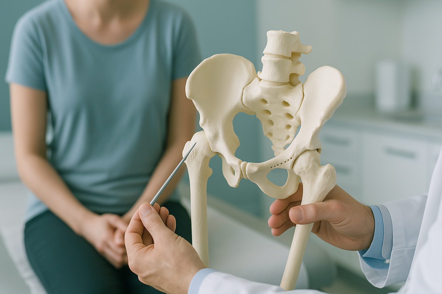
A periacetabular osteotomy (PAO) is a hip preservation operation for adults with hip dysplasia. The surgeon carefully cuts the hip socket and moves it to a new position, so it covers the ball of the thigh bone more fully. This improves joint stability, reduces pain, and can delay or prevent the need for a hip replacement later in life.
Key takeaways
- PAO moves the hip socket to cover the femoral head better, so you keep your own joint.
- It suits younger adults best, when the cartilage in the hip joint is still in reasonable condition.
- Accurate imaging of the pelvis under load before the operation helps the surgeon plan the correction.
- Recovery is gradual. It usually involves several months of protected weight-bearing.
- EOS standing imaging shows your pelvis and hip alignment as they truly are when you are upright.
How does a periacetabular osteotomy work?
A PAO changes the position of your hip socket (the acetabulum) so it sits more securely over the top of the thigh bone. In hip dysplasia in adults, the socket is too shallow. This means the ball of the hip joint is not held in properly. That leads to instability, labral tears and, in the end, arthritis.
During surgery, the surgeon makes precise cuts around the acetabulum and frees it from the rest of the pelvis. The surgeon then turns the socket into a better position and fixes it with screws. Your own bone and cartilage stay in place. That is why it is called a hip preservation procedure: it keeps your natural joint.
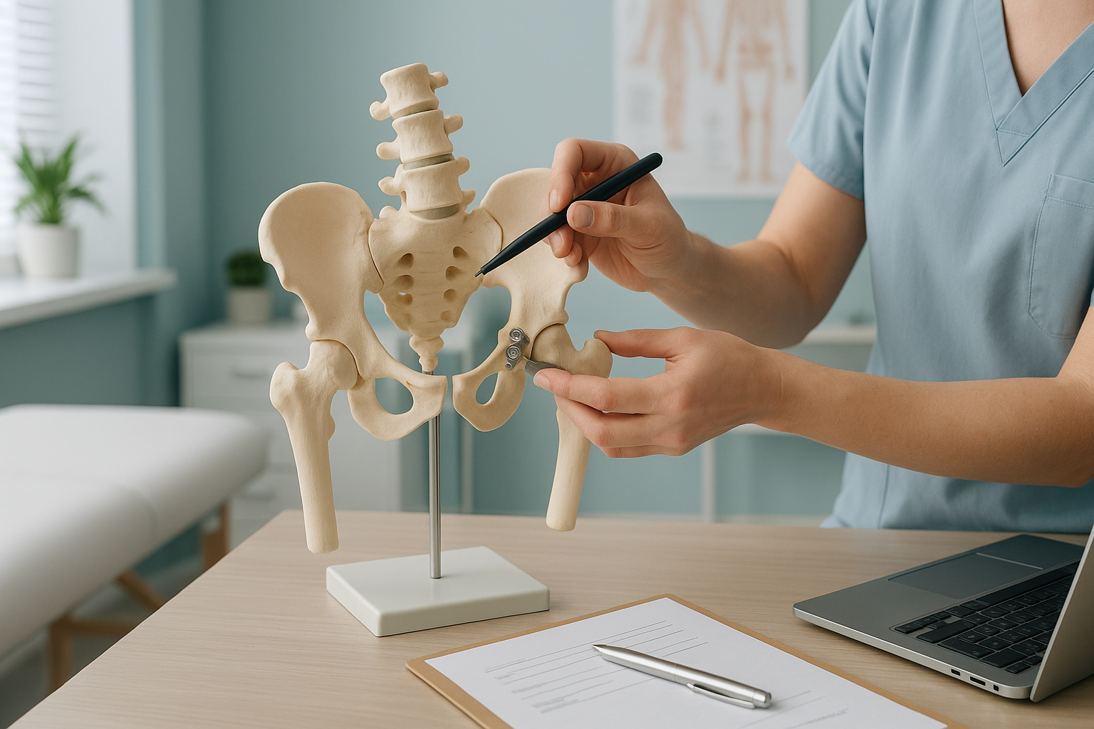
The operation is sometimes called a Ganz osteotomy, after the Swiss surgeon who developed the modern technique. It is a major procedure. But for the right patient it can be life-changing, giving years of better function on your own hip joint.
What symptoms suggest you might need a PAO?
Most people being considered for a periacetabular osteotomy have lived with hip dysplasia for years, sometimes without knowing it. Symptoms tend to build up slowly.
A deep ache in the groin or the front of the hip, often worse after walking or sitting for a long time.
A feeling that the hip catches or gives way. This may point to a labral tear caused by the instability.
Trouble keeping up with exercise, sport or daily activities that you used to manage without pain.
A tight feeling or limited movement, especially when turning the hip inward or bending deeply.
These symptoms can overlap with many other hip problems. That is why accurate imaging matters before any decision about surgery. The condition may be linked to developmental dysplasia of the hip, which sometimes is not picked up until adulthood.
Who is a good candidate for PAO surgery?
A PAO works best for adults whose hip cartilage is still in reasonable condition. The aim is to save the joint before arthritis sets in. It is not suitable for everyone.
- Young adults (typically under 40 to 50) with confirmed hip dysplasia
- Good remaining cartilage on imaging
- Pain or instability that is not getting better with physiotherapy
- Wanting to delay or avoid a hip replacement
- Significant cartilage loss or established arthritis
- Older adults, where a replacement may give a better outcome
- Other medical conditions that make major surgery risky
- Hip dysplasia that is very mild and can be managed without surgery
The decision is always made by a specialist hip preservation surgeon, not by an imaging clinic. ScanAlign provides the diagnostic images. Your surgeon reads them and advises you whether a PAO, a hip replacement or continued conservative management is the right path.
A note on MRI. EOS shows bones, joints and whole-body alignment under load. To assess cartilage, labral tears or soft tissue around the hip, MRI is usually the better tool. Your surgeon may ask for both types of imaging before planning a PAO.
Why does standing imaging matter before a PAO?
Standing imaging matters because your pelvis behaves differently when you are upright and carrying your full body weight than when you are lying on a scanning table. Standard X-rays and CT scans taken lying flat can relax the muscles. This lets the pelvis shift into a position it does not actually hold in daily life.
A PAO is all about moving the socket to the right angle. If the planning images show the pelvis in a relaxed, artificial position, the surgical plan may not match how you actually stand and move. EOS takes a full-length standing image of both hips and the entire pelvis in a single low-dose exposure, using about 90% less radiation than a standard X-ray, around ten times less.
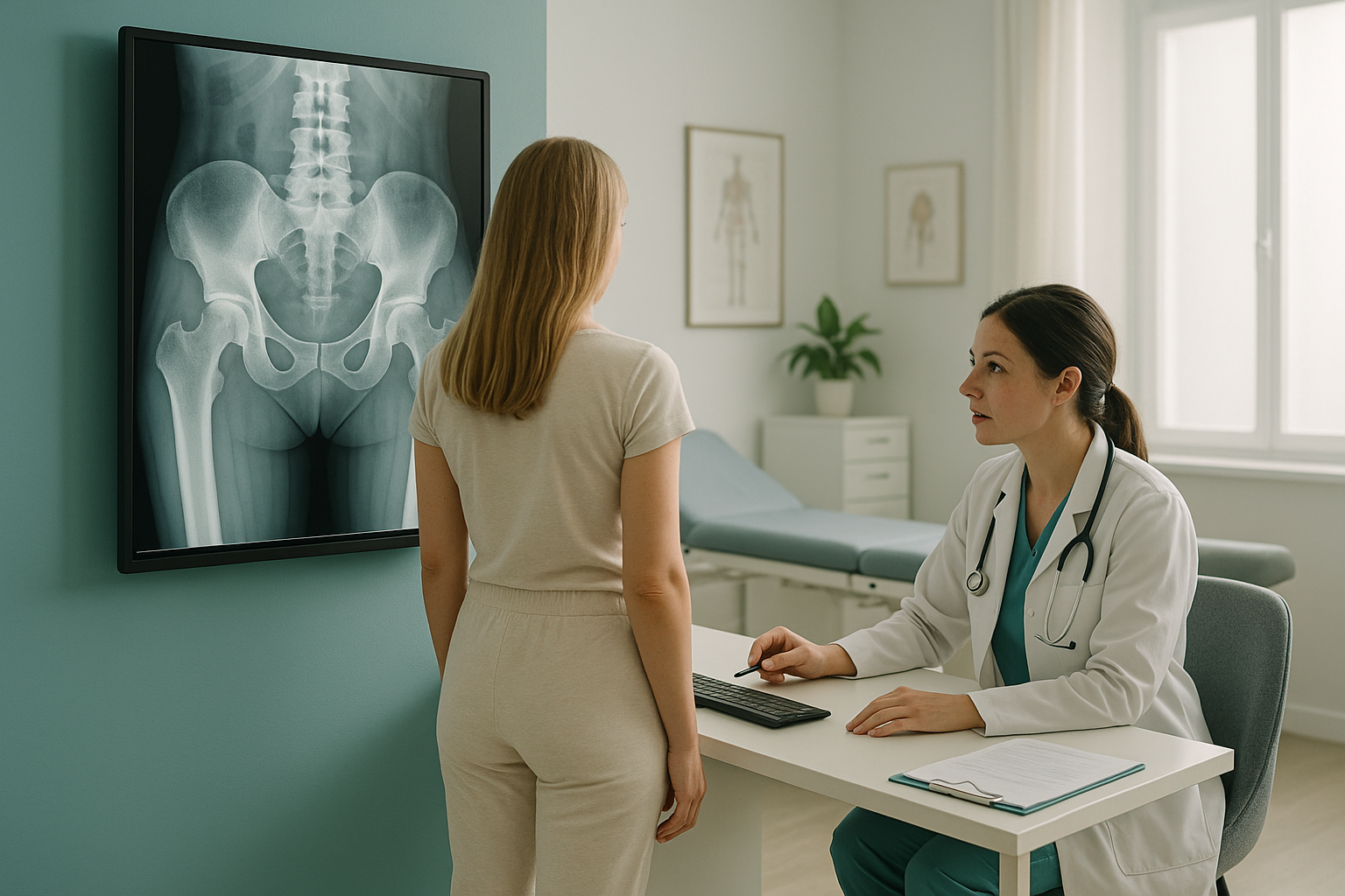
Your body is a connected chain. If your pelvis tilts forward or backward when you stand, the real coverage of the hip socket changes. A socket that looks adequate lying down might look inadequate standing up, or the other way round. For a procedure as precise as a PAO, that difference matters.
Thinking about PAO surgery and want standing pelvic imaging to support your surgical planning?
Book a free video consultationWhat does PAO recovery look like?
Recovery from a periacetabular osteotomy is gradual. It is a major operation with bone cuts and internal fixation, so your body needs time to heal properly.
First weeks
A hospital stay of several days, then protected weight-bearing with crutches. The focus is on wound healing and gentle range-of-motion exercises.
Weeks 6 to 12
A gradual return to weight-bearing as the bone heals. Physiotherapy becomes more active and builds strength around the hip.
Three to six months
Most people return to daily activities. Strengthening and mobility work continues. Follow-up imaging may be done to check bone healing.
Six months onward
A gradual return to sport or higher-impact activities, guided by your surgeon and physiotherapist. Full recovery can take nine to twelve months.
Every patient is different. Your surgeon and physiotherapy team will shape the timeline to your own situation. Standing imaging after surgery can show how the correction has changed your alignment under load. This gives your team a clear picture of how the hip is settling.
Before you start a treatment, or change the one you are on, it is worth getting a detailed diagnosis. Knowing whether the cause sits in the pelvis, the legs or the spine changes what will actually help.
Frequently asked questions about PAO surgery
It is an operation that moves the hip socket so it covers the ball of the thigh bone more completely. The surgeon cuts around the socket, turns it to a better angle and fixes it with screws. It keeps your own joint instead of replacing it.
PAO is most often done on younger adults, often in their twenties or thirties, because they tend to have better cartilage. However, some patients in their forties may still be suitable. The key factor is the condition of the cartilage, not just age alone.
Most people use crutches for around six to twelve weeks. A return to daily activities usually happens within three to six months. Full recovery, including higher-impact sport, can take nine to twelve months. Your surgeon will guide your own timeline.
A successful PAO can significantly delay the need for a hip replacement, sometimes by decades. It does not guarantee you will never need one. The outcome depends on factors such as the condition of the cartilage at the time of surgery and how active you are afterwards.
The pelvis tilts differently when you are standing than when you are lying down. That tilt changes how much the socket covers the femoral head. Standing imaging, such as EOS, shows the true position of the pelvis under load. This helps the surgeon plan the exact correction needed.
EOS uses about 90% less radiation than a standard X-ray, around ten times less. This makes it especially suitable when several images are needed before and after surgery.
No. At ScanAlign, standing EOS imaging in London, you can self-refer. Many patients take their images to their orthopaedic surgeon afterwards. You can also be referred directly by your consultant or physiotherapist.
For patients travelling from abroad, the price is confirmed during the free video consultation. We are a private, self-pay clinic.
Mild arthritis does not automatically rule out a PAO, but moderate to severe cartilage loss usually does. Your surgeon will assess your imaging carefully. In some cases, a hip replacement may be the more predictable option.
Sudden numbness, weakness, or loss of bladder or bowel control are red-flag symptoms. Do not wait for a scan. Contact your doctor or go to the nearest emergency department immediately for urgent medical attention.
Thinking about PAO surgery?
Standing pelvic imaging can give your surgeon a clearer picture of your hip alignment when you are carrying your real body weight. Start with a free video consultation to find out whether EOS imaging at 19 Harley Street, London, could support your surgical planning.
Book a free video consultationWritten by Abbas Dhami (Specialist Diagnostic Radiographer)
Sources
- Ganz R, Klaue K, Vinh TS, Mast JW. A new periacetabular osteotomy for the treatment of hip dysplasias. Clinical Orthopaedics and Related Research. 1988;(232):26-36.
- Clohisy JC, Schutz AL, St John L, Schoenecker PL, Wright RW. Periacetabular osteotomy: a systematic literature review. Clinical Orthopaedics and Related Research. 2009;467(8):2041-2052.
- International Hip Dysplasia Institute. Adult Hip Dysplasia: Information for Patients. hipdysplasia.org.
This page is general information and not a diagnosis.
ScanAlign is the trading name of CoreMed Solutions Ltd and operates under The Harley Street Hospital's CQC licence.