
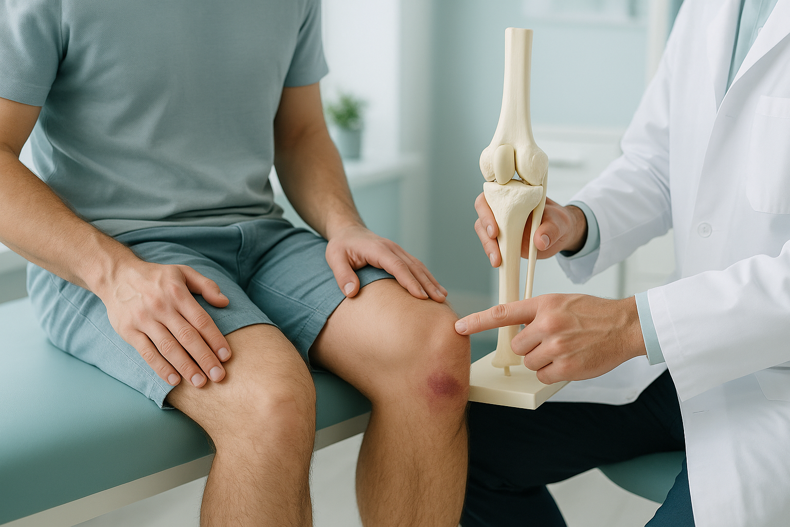
What Is a Patellar Tendon Rupture, and How Is It Repaired?
A patellar tendon rupture is a complete tear of the tendon that joins your kneecap to your shinbone. It breaks the extensor mechanism of the knee. This makes it impossible to straighten your leg under your own power. It is an urgent injury that almost always needs surgical repair. If you think you have a rupture, go to a hospital emergency department straight away.
Key takeaways
- A ruptured patellar tendon is an emergency. You should get urgent medical help, not book a scan.
- Surgery to reattach the tendon is usually needed within days of the injury.
- Recovery usually takes many months of structured rehabilitation.
- After repair, standing whole-body imaging can help your surgeon check the position of the kneecap (patella) and leg alignment during recovery.
- The patellar tendon is different from the quadriceps tendon. The quadriceps tendon sits above the kneecap, not below it.
Urgent. A patellar tendon rupture is a serious acute injury. If you have sudden knee pain, a popping feeling, swelling, and you cannot straighten your leg, go to a hospital emergency department immediately. Do not wait to book a private scan. ScanAlign does not assess or treat acute injuries. Our imaging is for follow-up and recovery assessment only.
What are the symptoms of a patellar tendon rupture?
The most obvious symptom is that you cannot straighten your knee after the injury. You may have felt or heard a pop at the front of the knee. The kneecap often rides higher than normal, because nothing is holding it down from below.
You may also see a gap or dent just below the kneecap, where the tendon has torn away. Walking becomes very difficult or impossible.
What causes a patellar tendon to rupture?
The most common cause is a sudden, forceful tightening of the quadriceps muscle while the knee is bent. Examples are landing from a jump or stumbling off a step. The tendon gives way under the load.
The rupture usually happens where the tendon attaches to the bottom of the kneecap. In some cases it tears away from where it attaches on the shinbone instead.
How does the extensor mechanism of the knee work?
The extensor mechanism is the chain of structures that lets you straighten your knee. The quadriceps muscle joins the top of the kneecap through the quadriceps tendon. The patellar tendon then joins the bottom of the kneecap to the tibial tuberosity, a bony bump at the top of your shinbone. If either tendon ruptures, the chain is broken and the knee cannot straighten.
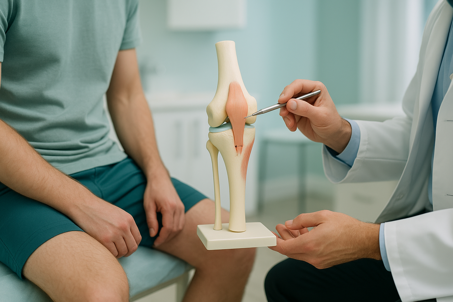
A quadriceps tendon rupture breaks the chain above the kneecap. A patella ligament rupture breaks it below. Both stop you from straightening your leg, but the surgical approach is different.
How is a torn patellar tendon repaired with surgery?
Surgical repair is almost always necessary. The torn ends of the tendon cannot heal on their own, because the quadriceps muscle pulls them apart. For the best results, surgery is usually done within the first week or two after the injury.
What happens during the operation
The surgeon makes a cut over the front of the knee. They reattach the tendon to the kneecap with strong stitches passed through holes drilled in the bone. In some cases, a wire or cable is placed for a while to protect the repair as it heals. If the tendon tissue is too damaged, a graft from another tendon may be used to strengthen the repair.
The aim is to bring the patella back to its correct position, so the extensor mechanism works properly again. If the kneecap sits too high after repair, the knee may not bend or straighten normally.
What does recovery after patellar tendon repair look like?
Recovery is long and structured. Most people spend several months in a rehabilitation programme. It slowly builds quadriceps strength and brings back the knee's range of movement.
Immobilisation (weeks 0 to 6)
The knee is held in a brace, usually locked straight, to protect the repair. Putting weight on the leg may be limited.
Early movement (weeks 6 to 12)
Gentle bending starts with guidance from a physiotherapist. The brace may be unlocked step by step.
Strengthening (months 3 to 6)
Focused quadriceps strengthening exercises rebuild muscle power and knee stability.
Return to activity (months 6 to 12+)
Going back to sport depends on strength recovery, tendon healing and alignment. Some people take a full year.
Throughout this process, your surgical team will want to check that the patella is sitting in the right position and that leg alignment is on track.
How does standing imaging help after patellar tendon repair?
Standard X-rays are usually taken lying down. That position relaxes the muscles and removes the effect of gravity. So the kneecap may look normal even when it does not sit normally under real-life load. A standing, weight-bearing image shows where the patella actually sits when you are on your feet.
ScanAlign standing EOS imaging takes a full-length image from your hips to your ankles in one single exposure, with about 90% less radiation than a standard X-ray, around ten times less. This lets your surgeon see the patella position in relation to the whole leg. That includes any changes in knee alignment or leg length that may have developed during recovery.
Your body is a connected chain. When one knee is kept still for weeks, the hip, ankle and other leg often compensate. A whole-body standing image shows these compensations, so they can be addressed during rehabilitation.
If your surgeon wants to check patella position and leg alignment after your repair, you can book follow-up alignment imaging at ScanAlign in London.
Book a free video consultationWhen MRI is the better tool. EOS imaging shows bones, joints and alignment. For soft-tissue detail, such as the tendon itself, nearby ligaments or cartilage damage, MRI is usually the better option. Your consultant will advise which imaging suits your stage of recovery.
Is a standing alignment scan right for you?
ScanAlign does not assess or treat acute ruptures. Our imaging is designed for the recovery phase. This is when you and your surgeon want to understand alignment and patella position under real weight-bearing conditions.
Before you start a treatment, or change the one you are on, it is worth getting a detailed diagnosis. Knowing whether the cause sits in the pelvis, the legs or the spine changes what will actually help.
- You have had patellar tendon repair and want to check patella height
- Your surgeon wants a standing full-leg alignment view
- You are months into recovery and your knee still does not feel right
- You have just injured your knee and cannot put weight on it. Go to a hospital emergency department.
- You need soft-tissue or tendon imaging. Ask about MRI instead.
- You have sudden numbness, weakness, or loss of bladder or bowel control. Get urgent medical help.
Frequently asked questions
In almost all cases, no. The quadriceps muscle pulls the torn ends apart, so the tendon cannot join back on its own. Surgical reattachment is the standard treatment. Only in very rare cases, such as when a patient is too unwell for surgery, might treatment without surgery be tried.
Both break the extensor mechanism of the knee. The patellar tendon sits below the kneecap and joins it to the shinbone. The quadriceps tendon sits above and joins the thigh muscle to the kneecap. Both injuries stop you from straightening the knee. But patellar tendon ruptures are more common in younger, active adults, while quadriceps tendon ruptures tend to happen in people over 40.
Full recovery usually takes six months to a year. The first six weeks are spent in a brace with limited movement. Physiotherapy then moves on to range-of-movement exercises and strengthening. Going back to sport or heavy activity usually takes at least nine to twelve months, depending on progress.
Patella alta means the kneecap is sitting higher than normal. After a patellar tendon rupture, the quadriceps muscle pulls the kneecap upward, because the tendon below is no longer holding it down. Patella alta can also develop after repair if the tendon heals in a lengthened position. Standing imaging helps assess patella height under weight-bearing conditions.
If you cannot actively straighten your knee, the extensor mechanism may be disrupted. This could be a patellar tendon rupture, a quadriceps tendon rupture or a patella fracture. This is an urgent finding. Please go to a hospital emergency department for assessment straight away.
Yes. Chronic patellar tendonitis causes ongoing micro-damage inside the tendon and weakens it over time. A tendon that has been inflamed or degenerated for months is more vulnerable to a sudden complete tear, especially during explosive movements.
An EOS scan takes a full-length, weight-bearing image of your legs while you are standing. Standard X-rays are taken lying down, which relaxes the muscles and removes the effect of gravity. A standing image shows real-life patella position and leg alignment. This gives your surgeon a more accurate picture.
Yes. EOS uses about 90% less radiation than a standard X-ray, around ten times less. It is widely used in orthopaedic assessment and is considered very safe for adults.
No. You can book yourself. The process starts with a free video consultation. A specialist radiographer reviews your situation and confirms whether EOS imaging is right for you.
ScanAlign is a private, self-pay clinic. The price for patients travelling from abroad is confirmed during the free video consultation.
Recovering from patellar tendon repair?
If you want to check your patella position and leg alignment during recovery, start with a free video consultation with our specialist radiographer. You can do it from home before you plan any travel to London.
Book a free video consultationWritten by Abbas Dhami (Specialist Diagnostic Radiographer)
Sources
- Saragaglia D, et al. "Patellar tendon rupture: results of surgical treatment." Revue de Chirurgie Orthopédique et Réparatrice de l'Appareil Moteur. Published by Elsevier Masson.
- NHS. "Tendon injuries." NHS Inform. Available at nhs.uk.
- American Academy of Orthopaedic Surgeons (AAOS). "Patellar Tendon Tear." OrthoInfo patient education resource. Available at orthoinfo.aaos.org.
This page is general information and not a diagnosis.
ScanAlign is the trading name of CoreMed Solutions Ltd and operates under The Harley Street Hospital's CQC licence.