
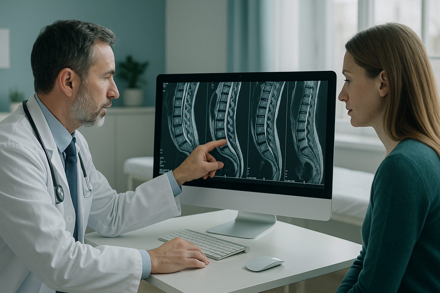
When Should a Pain Specialist Look Beyond a Normal Scan?
Sometimes chronic pain carries on even though the MRI or CT scan is normal. In these cases, standing whole-body imaging can show alignment problems that depend on load. These problems only appear under the patient's own body weight. Scans done lying down relax the muscles and take gravity away, so they may hide the structural cause. A second look while the patient bears weight does not replace MRI. It adds a different piece of the diagnostic picture, one that is sometimes missing.
Key points
- A "normal" scan done lying down does not rule out a postural or load-dependent structural cause.
- Pain that gets worse with standing, walking or holding one posture for a long time is a clinical clue. It is worth checking with the patient upright.
- Standing whole-body EOS imaging captures alignment from head to feet in a single exposure, with about 90% less radiation than a standard X-ray.
- Many cases of chronic pain are not explained by any structural finding. Imaging should only be requested when there is a clear clinical question.
- ScanAlign shares its reports, so the findings fit into the pain management plan already in place.
Why does a normal MRI not always explain chronic pain?
A normal MRI means the scan did not find a lesion, disc herniation, tumour or inflammatory process in the area scanned. It does not mean nothing structural is adding to the patient's pain. MRI and CT are done lying down. In that position, the spine decompresses and the pelvis settles into a neutral position. The muscles that normally hold the body upright are relaxed. Any alignment problem that depends on gravity and load is, in effect, switched off during the scan.
This matters because the patient's pain usually happens when they are upright: walking, standing or sitting. If the problem is positional, the image taken lying down may look completely normal. The scan answered one question well, but a different question was never asked.
To learn more about this gap, read about when an MRI shows nothing but the pain remains.
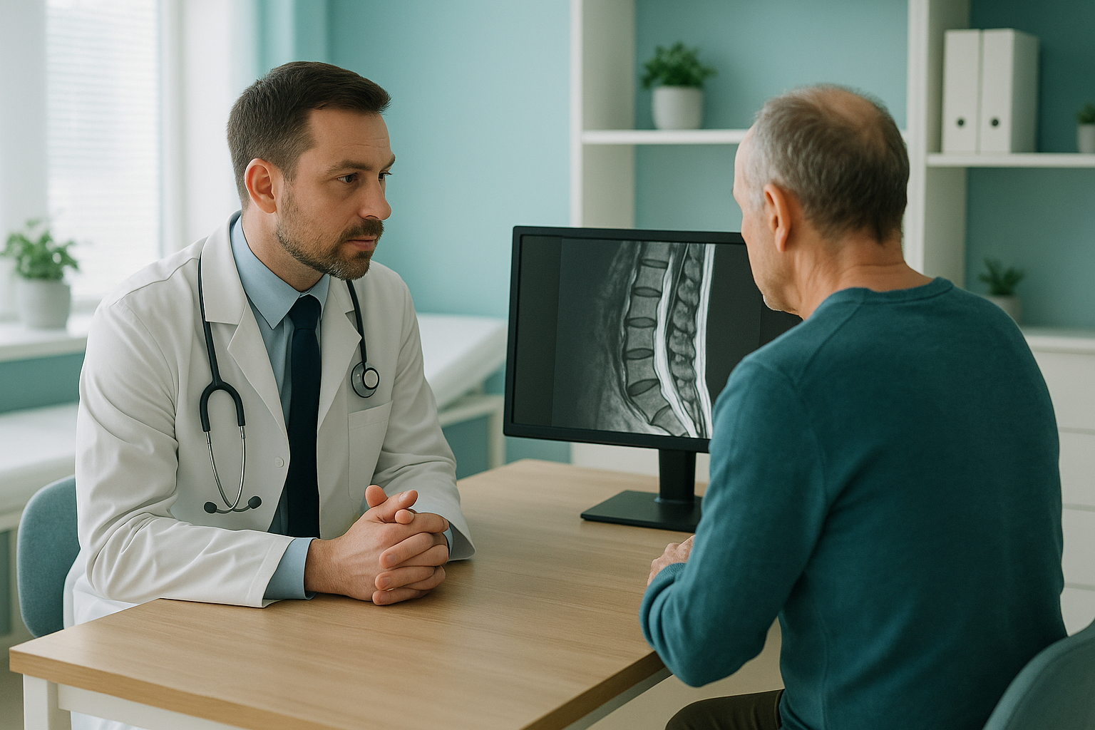
What clinical clues suggest a load-dependent structural cause?
Some patterns in the history and examination make it more likely that a postural or biomechanical factor is adding to ongoing pain. They do not make a diagnosis on their own. But they point to a question that imaging done lying down cannot answer.
Pain that gets worse with standing or walking, and gets better when the patient lies down or changes position.
Knee, hip or thoracic pain that does not match a single structural lesion. This suggests a compensation pattern further along the chain.
A leg length difference, lateral trunk shift or pelvic obliquity that can be seen on clinical examination.
Pain that carries on after spinal, hip or knee surgery, where imaging of that area looks satisfactory but overall alignment was never assessed.
The body is a connected chain from feet to head. A problem at one level can make another area compensate. This means the place of the pain is not always the place of the cause. Imaging of one area shows that area well. Whole-body standing imaging shows how everything lines up together.
Before a patient starts a treatment, or changes the one they are on, it is worth getting a detailed diagnosis. Knowing whether the cause sits in the pelvis, the legs or the spine changes what will actually help.
How does standing whole-body imaging differ from usual scans?
Standing whole-body EOS imaging captures the full skeleton from head to feet in a single exposure, while the patient bears weight. Because the patient is upright, the image reflects the real mechanical setting in which their pain happens. Gravity, muscle tone and compensation patterns are all present in the image, instead of being removed by lying flat on the back.
What it shows: A detailed view of one body area (for example the lumbar spine or a single knee). Excellent for finding disc herniations, fractures, tumours and inflammatory changes in that area.
What it may miss: How that area relates to the rest of the skeleton under load. A "normal" lumbar MRI cannot tell you whether the pelvis is tilted, the legs are unequal, or the thoracic spine is compensating.
What it shows: Full skeletal alignment from head to feet, all in one standing image. Pelvic obliquity, sagittal balance, limb length difference and compensatory curves can all be seen and measured.
What it does not show: Soft tissue detail. It will not find a disc herniation, nerve root compression, ligament tear or inflammatory process. For those questions, MRI remains the right tool.
EOS uses about 90% less radiation than a standard X-ray, around ten times less. That lower dose makes full-body imaging practical, without the radiation burden of stitched full-spine plain films. A detailed alignment report with measurements is provided. It is designed to sit alongside existing MRI or CT findings, so the treating team has the full picture.
When will more imaging not help a patient with chronic pain?
Not every patient with unexplained pain has a structural cause waiting to be found. It is important to be clear about the limits of any type of imaging, including standing whole-body imaging.
- Pain is clearly linked to position or load
- Clinical examination suggests asymmetry or postural imbalance
- Imaging of the area is normal but a structural question remains
- Planning before surgery needs the full alignment picture
- Pain after surgery carries on despite satisfactory imaging of the area
- Pain is mainly driven by central sensitisation, with no likely structural factor
- The clinical picture is mainly psychological or social
- Repeated normal imaging has already been discussed and the patient needs reassurance, not more scans
- Red flags (neurological deficit, unexplained weight loss, suspected malignancy) need an urgent MRI or other targeted investigation
Claiming too much for any scan risks strengthening unhelpful beliefs about illness. Standing alignment imaging is one extra tool. It answers a specific structural question that imaging done lying down cannot. But it is not right for every case of chronic pain.
A note on MRI: EOS shows bones, joints and alignment under load. For soft tissue, discs or nerves, MRI is usually the better tool. The two work together. An EOS alignment report works best when read alongside existing MRI or CT findings.
How can a pain specialist refer a patient for standing imaging?
Referring a patient to ScanAlign, whole-body standing imaging at 19 Harley Street, London, is simple. The service is self-pay, so no insurance pre-approval is needed, though some insurers do cover the scan.
Register your clinic
Fill in a short form to register to refer patients. It only takes a moment and lets us share reports directly with you.
Send a referral
Once registered, you can refer a patient online. Include any relevant clinical history or imaging already done.
The patient comes to London
The scan is done standing up. It captures the full skeleton in a single low-dose exposure. There is no contrast injection and no enclosed scanner.
Shared report
A detailed alignment report with measurements is sent to you and the patient. The findings are designed to fit into your existing management plan.
Patients can also refer themselves directly. They start with a free video consultation to check the scan is right for their situation.
Book a free video consultationFrequently asked questions
EOS is a low-dose standing X-ray system. It captures the full skeleton from head to feet in one exposure. Usual X-rays image a single area lying down. EOS instead produces a whole-body image while the patient bears weight, with about 90% less radiation than a standard X-ray, around ten times less.
MRI is done lying down, which removes gravity and relaxes the muscles. Alignment problems that depend on load, such as pelvic obliquity, sagittal imbalance or functional leg length difference, may not be visible in that position. The MRI answered its question well, but the question about position was never asked.
No. It is most useful when there is a clinical suspicion of a load-dependent structural factor. Some patients are unlikely to benefit: those whose pain is mainly driven by central sensitisation or psychological factors, or those for whom repeated normal imaging would strengthen unhelpful beliefs.
No. EOS shows bones, joints and skeletal alignment. For soft tissue detail, disc herniations or nerve root compression, MRI remains the right investigation. The two work together.
EOS uses about 90% less radiation than a standard X-ray, around ten times less. This makes full-body imaging practical, without the build-up of dose that comes with a stitched series of plain films.
Register your clinic online, then send referrals through a short form. Patients can also refer themselves and start with a free video consultation to check the scan is suitable. The service is self-pay, though some insurers cover the scan.
For patients travelling from abroad, the price is confirmed during the free video consultation. The service is self-pay, and some patients may be able to claim through their health insurer.
Yes. A detailed alignment report with measurements is produced and shared with both you and the patient. It is designed to sit alongside existing MRI or CT findings, giving you the full structural picture.
Yes. No referral from their doctor or a specialist is needed. Patients can book a free video consultation directly to discuss whether standing imaging is right for their situation.
ScanAlign is at 19 Harley Street, London, within The Harley Street Hospital. The clinic operates under the hospital's CQC licence.
A second look, standing up
When imaging done lying down leaves a structural question unanswered, standing whole-body EOS imaging can give the missing context for your pain management plan.
Book a free video consultationWritten by Abbas Dhami (Specialist Diagnostic Radiographer)
Sources
- Illés, T. & Somoskeöy, S. (2012). The EOS imaging system and its uses in daily orthopaedic practice. International Orthopaedics, 36(7), 1405-1411.
- Roussouly, P. & Pinheiro-Franco, J.L. (2011). Biomechanical analysis of the spino-pelvic organisation and adaptation in pathology. European Spine Journal, 20(Suppl 5), 609-618.
- National Institute for Health and Care Excellence (NICE). Low back pain and sciatica in over 16s: assessment and management (NG59).
This page is general information and not a diagnosis.
ScanAlign is the trading name of CoreMed Solutions Ltd and operates under The Harley Street Hospital's CQC licence.