
How Does Osteoporosis Change the Shape of Your Spine?
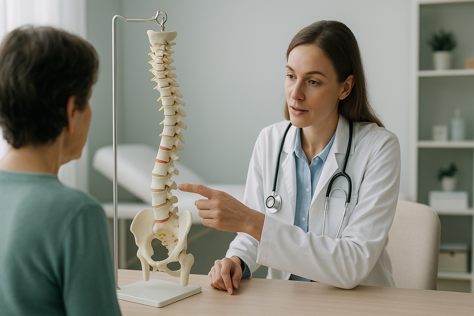
Osteoporosis weakens the vertebrae, so they can slowly collapse under your own body weight. These small compression fractures turn single bones into wedges. This pulls the spine forward into a rounded curve called kyphosis. Over time, this changes your posture and reduces your height. It also shifts your centre of gravity and can cause pain in areas far away from the fracture itself.
Key points
- Vertebrae weakened by osteoporosis can break silently, without a fall or clear injury.
- Each wedge fracture tips the spine further forward. The curve gets bigger and you lose height.
- A standing scan shows the real shape of the spine under gravity. Scans done lying down can hide this.
- EOS does not measure bone density. A DEXA scan is the correct test for that.
- Knowing your full alignment helps clinicians plan the right support and prevent further collapse.
What does osteoporosis actually do to the spine?
Osteoporosis makes bones lose density and strength. When the vertebrae become porous and fragile, they can no longer carry your body weight in the same way. The front edge of a vertebra may squash down more than the back. The bone then changes from a rectangular block into a wedge shape. Doctors call this a wedge fracture or compression fracture.
One wedge fracture tilts the vertebra above it slightly forward. If several vertebrae collapse like this, the effect adds up. The upper back rounds into an exaggerated forward curve. This condition is called kyphosis. You may know it as a stooped posture, sometimes called dowager's hump.

The spine works as a connected chain, so a change in one area forces everything else to adjust. Your hips may tilt and your head may push forward. Muscles all through your back and legs work harder just to keep you upright. This is why pain from osteoporosis changes in the spine often shows up in places you would not expect, such as the lower back, the hips or even the knees.
What are the signs that osteoporosis is affecting your spine?
Many compression fractures happen without any dramatic event. You might not remember a specific injury. Instead, the signs tend to creep in slowly over months or years.
Losing more than a couple of centimetres over the years can point to collapse of the vertebrae rather than normal ageing.
An upper back that grows more and more rounded, making it hard to stand fully upright, even when you try.
A dull, nagging pain between the shoulder blades that gets worse with standing or walking.
As the spine curves forward, your centre of gravity shifts. This makes you less steady on your feet.
Important: if you have sudden, severe back pain after a minor bump or fall, or any new numbness, weakness, or loss of bladder or bowel control, get urgent medical help from your doctor or your nearest emergency department. These symptoms need assessment straight away, not an imaging booking.
What causes osteoporosis in the first place?
Bone is living tissue that is always rebuilding itself. Osteoporosis develops when the body breaks down old bone faster than it can replace it. This leaves a weaker framework behind.
Bone density naturally peaks in your thirties and falls with age, especially after the menopause.
Lower oestrogen or testosterone levels speed up bone loss significantly.
Without the right nutrients, your body cannot keep a strong bone structure.
Long-term steroid use and certain medical conditions can weaken bones faster.
It is worth knowing that osteoporosis and osteomalacia are different conditions. Osteoporosis means the bone structure is thinning. Osteomalacia means the bone is poorly mineralised, often because of severe vitamin D deficiency. Both can affect the spine, but they need different tests and management.
Why does a standing scan show spine changes that a lying-down scan might miss?
A standing scan captures your spine exactly as it works in real life, under the full pull of gravity. When you lie down for a usual X-ray, CT or MRI, your muscles relax and the spine straightens out. A forward curve that is very clear when you are upright can look much less severe when you are flat on a table.
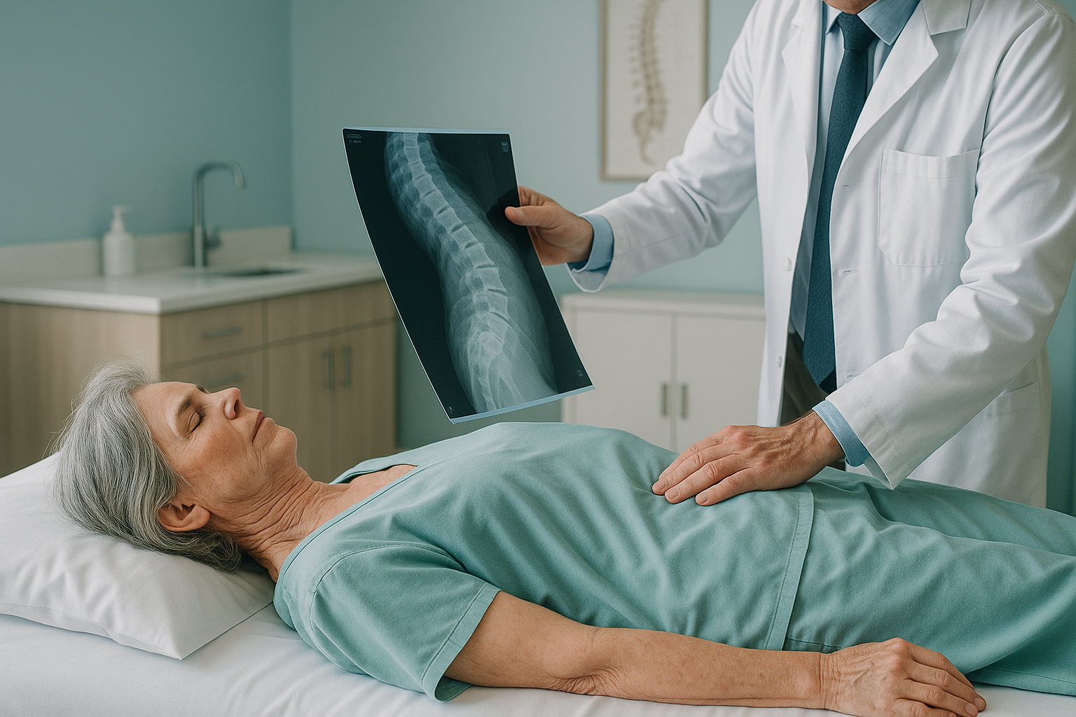
This matters because your specialist's decisions about treatment, bracing or exercise all depend on how your spine really behaves when you are upright. ScanAlign, low dose standing imaging uses EOS technology to capture your full skeleton from head to feet in a single standing image, using about 90% less radiation than a standard X-ray, around ten times less.
An EOS scan shows the overall sagittal balance of your body. This means your specialist can see not just the curve in your upper back, but also how your pelvis, hips and legs are making up for it. Imaging of a single area misses this whole-body view.
EOS and bone density: an EOS scan shows the shape and alignment of your bones and joints. It does not measure bone density. If your doctor needs to know how porous your bones are, a DEXA scan is the right test for that. The two work well together: DEXA tells you how strong the bone is, and EOS tells you how the skeleton is lined up as a result.
When MRI is the better choice: EOS is excellent at showing bone alignment and joint positions. For soft-tissue detail, such as discs, nerves or the spinal cord itself, MRI is usually the better tool. Your clinician can advise which scan, or which combination, suits your situation.
What can be done once osteoporosis has changed the spine?
How osteoporosis changes in the spine are managed depends entirely on how far the condition has progressed. It also depends on how it affects your balance, comfort and daily life. ScanAlign is a diagnostic imaging clinic, so we do not provide treatment. But understanding your alignment is often the first step that lets your medical team plan the right approach.
Common options your clinician might consider include:
- Medicine to slow further bone loss and reduce the risk of new fractures.
- Physiotherapy and targeted exercise to strengthen the muscles that support the spine and improve balance.
- Bracing in some cases, to support the spine while fractures heal.
- Procedures such as kyphoplasty or vertebroplasty for painful compression fractures that have not responded to other measures.
Accurate alignment imaging helps your team choose between these options. For example, knowing how far forward your centre of gravity has shifted determines whether a brace alone might help, or whether a more active treatment is worth discussing.
It is also worth knowing that not every rounded spine in older age is caused by osteoporosis. Scheuermann's kyphosis, for example, has a different underlying cause and needs a different approach. A standing whole-body image helps tell these conditions apart by showing the exact shape and position of each vertebra under load.
Before you start treatment, or change the one you are on, it is worth getting a detailed diagnosis. Knowing whether the cause sits in the pelvis, the legs or the spine changes what will actually help.
Wondering whether your posture changes are linked to bone health? A free video consultation lets you talk through your situation with our specialist radiographer before you decide on any imaging.
Book a free video consultationFrequently asked questions
Yes. Each compression fracture in the spine takes away a small amount of height. After several fractures, you can lose noticeable height, sometimes several centimetres over the years.
No. EOS shows the shape and alignment of your bones, not their density. A DEXA scan is the standard test for measuring bone density. The two types of scan work well together.
An EOS scan uses about 90% less radiation than a standard X-ray, around ten times less. This makes it especially suitable when imaging the full spine and legs in a single session.
When you lie down, gravity no longer pulls on the spine, so curves can flatten and look less severe. A standing scan captures your real alignment under load. This gives your specialist the truest picture of how your posture has changed.
Yes. In bone weakened by osteoporosis, everyday activities like bending, lifting a bag or even coughing can be enough to make a vertebra fracture. Many people do not know that a fracture has happened.
No. ScanAlign accepts self-referrals. You can book a free video consultation to discuss whether imaging is right for your situation, without needing a letter from your doctor.
The price for patients travelling from abroad is confirmed during the free video consultation. The consultation is also a good first step to understand whether a scan would be helpful for you.
Dowager's hump is an older, informal term for the rounded upper back (kyphosis) that often results from compression fractures caused by osteoporosis. Both terms describe the same visible change, though kyphosis can also have other causes.
Sagittal balance describes how well your body is centred over your pelvis when seen from the side. Good sagittal balance means you can stand comfortably without too much muscle effort. When osteoporosis pushes the spine forward, the whole body has to make up for it. This increases strain, tiredness and the risk of falls.
Once a vertebra has collapsed, the change in shape is generally permanent. However, treatment can prevent further fractures, improve muscle support around the spine and reduce pain. Accurate alignment imaging helps your medical team decide on the most suitable next steps.
See how your spine is really aligned
A free video consultation with our specialist radiographer helps you understand whether standing whole-body imaging could give you the answers you have been looking for.
Book a free video consultationWritten by Abbas Dhami (Specialist Diagnostic Radiographer)
Sources
- NHS. Osteoporosis: Overview. nhs.uk/conditions/osteoporosis (accessed 2024).
- Royal Osteoporosis Society. Vertebral fractures (spinal fractures). theros.org.uk (accessed 2024).
- Glassman SD, Bridwell K, Dimar JR, et al. The impact of positive sagittal balance in adult spinal deformity. Spine. 2005;30(18):2024-2029.
This page is general information and not a diagnosis.
ScanAlign is the trading name of CoreMed Solutions Ltd and operates under The Harley Street Hospital's CQC licence.