

What Is Osteochondritis Dissecans, and Who Gets It?
Osteochondritis dissecans (OCD) is a condition in which a small area of bone just under the cartilage surface of the knee loses its blood supply. This bone becomes soft. The cartilage above it may then loosen or break off, forming a loose body in the joint. It is most common in physically active adults and younger athletes.
Key points
- OCD affects the subchondral bone, the layer directly beneath cartilage. It is usually on the inner side of the knee.
- Symptoms include deep knee pain, swelling, and sometimes catching or locking if a fragment comes loose.
- MRI is the main test for staging the lesion. Standing imaging can add alignment details that lying-down scans miss.
- Early diagnosis matters. Smaller, stable lesions often respond to rest and changes in activity.
- A standing, weight-bearing scan shows how the alignment of your whole leg may be loading the affected area unevenly.
You may have been told you have osteochondritis dissecans. Or your knee may ache after sport and sometimes catch. This page explains what is happening inside your joint, and where standing imaging fits in.
What does osteochondritis dissecans feel like?
The first sign is usually a vague, deep ache inside the knee. It gets worse with activity, especially running, jumping or climbing stairs. As the condition progresses, you may notice more specific symptoms.
A dull ache deep inside the knee, usually on the inner side. It flares up during and after activity.
The knee may swell up after exercise and take hours or days to settle.
If a piece of cartilage loosens, it can get caught between the joint surfaces. This can make the knee lock or give way suddenly.
The knee may feel stiff after sitting. Over time, the thigh muscles can weaken because they are used less.
Important: if your knee locks completely and you cannot straighten it, or you have sudden severe swelling, see your doctor or go to a hospital emergency department promptly. A locked knee from a loose body may need urgent assessment.
What causes osteochondritis dissecans in the knee?
The exact cause is not fully understood. It involves a disruption of blood flow to a small area of subchondral bone, the hard layer directly beneath the joint cartilage. Without enough blood supply, that patch of bone weakens. Several factors are thought to play a part.
Repeated loading from running, jumping or pivoting sports can damage the blood vessels that feed the bone beneath cartilage.
Some people may naturally have a more fragile blood supply to certain parts of the knee bone. This makes those spots more prone to damage.
If your leg is angled inward or outward, some parts of the knee surface carry more weight than they should. This uneven loading can focus stress on one area of bone.
Most OCD lesions occur on the medial femoral condyle, the rounded bump on the inner side of the thighbone where it meets the knee. This area bears the most load when you walk, run and land from jumps.

How is osteochondritis dissecans diagnosed?
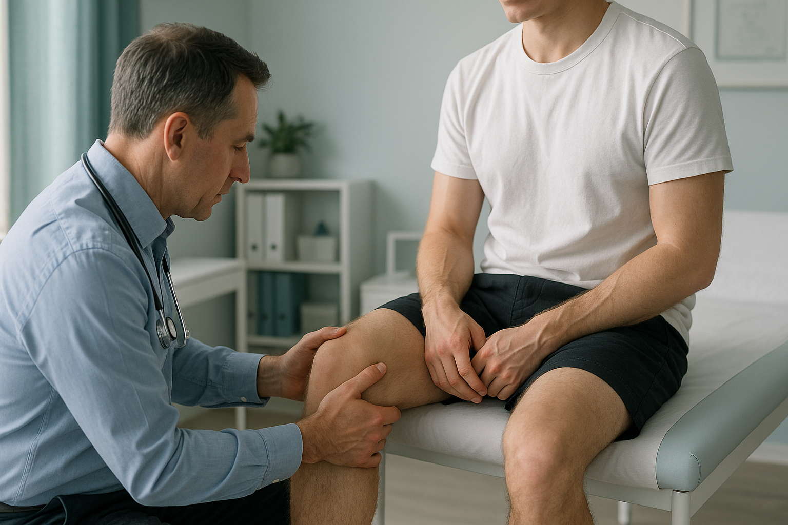
OCD is usually diagnosed with a clinical examination together with imaging. Your doctor will ask about your symptoms and check for tenderness on the inner knee. They may also check for swelling or limited movement.
Standard X-rays can sometimes show an OCD lesion. But they are taken lying down and only give a flat, two-dimensional view. MRI is the key test for staging the lesion. It shows whether the cartilage cap is intact, how large the affected area of bone is, and whether a fragment is at risk of separating.
When is MRI the right choice? For osteochondritis dissecans, MRI is usually the better test for seeing the cartilage surface, the bone beneath it, and any fluid signals that suggest the fragment is loosening. EOS standing imaging does not replace MRI for staging OCD. What it does is add alignment details. It shows how your full leg loads the knee when you are upright, which a lying-down MRI cannot capture.
Why does leg alignment matter if you have an OCD lesion?
Because the way your leg is aligned changes where force builds up across your knee. When you lie down for an MRI or standard X-ray, your muscles relax and gravity stops loading the joint. Alignment problems that only appear under your body weight are, in effect, hidden.
Your body works as a connected chain, from your foot through your ankle, knee, hip and spine. If your leg angles slightly inward, more force falls on the inner knee. This is exactly where most OCD lesions develop. A tilt at the hip or foot can shift loading higher up or lower down the chain. The knee then compensates in ways that focus stress on a small area of bone.
A standing, full-leg EOS scan at ScanAlign, low dose standing imaging captures your whole leg from hip to ankle in one image while you bear weight naturally. It uses about 90% less radiation than a standard X-ray, around ten times less. The result gives your specialist a clear picture of your leg alignment in real-life conditions. This information helps guide whether activity changes, bracing or surgical planning is most suitable.
What are the options if you have osteochondritis dissecans?
Treatment depends on the size and stability of the lesion, your age and how active you are. ScanAlign is a diagnostic imaging clinic, not a treatment centre, so we do not provide treatment. What we can do is give you and your specialist the alignment information that helps shape the right plan.
In general, the options your doctor may discuss include:
- Changing activity and rest. Cutting down on impact sports so the bone can heal. This is often tried first for stable lesions.
- Physiotherapy. Strengthening the muscles around the knee to support the joint and reduce loading on the affected area.
- Surgery. If the fragment has loosened or come away, surgical options may include fixing it back in place. If that is not possible, the loose body may be removed and the cartilage surface restored. Alignment correction may also be considered if the shape of the leg is adding to uneven loading.
Knowing how your leg is aligned under weight helps your surgeon decide whether a straightforward cartilage procedure is enough. Or whether the underlying alignment needs treating at the same time, to protect the repair in the long term.
If you are thinking about surgery, or want your specialist to see your full leg alignment, you can book a standing knee scan in London and take the results to your consultation.
Book a free video consultationIs a standing alignment scan right for you?
A standing EOS scan does not replace MRI for staging your OCD lesion. It adds something different: a view of your whole leg alignment under real body weight. Here is a quick guide.
- Have been diagnosed with OCD and want alignment details for your specialist
- Are being considered for surgery and need full-leg measurements
- Have knee pain that has not improved with rest and want to rule out alignment as a factor
- Want a very low-radiation image of both legs for comparison
- Need to see the cartilage surface and bone detail of the lesion itself
- Have soft-tissue symptoms, such as ligament or meniscus concerns
- Need to stage an OCD lesion for the first time
For younger patients, paediatric imaging at ScanAlign follows specific protocols designed to keep radiation as low as possible.
Before you start treatment, or change the one you are on, it is worth getting a detailed diagnosis. Knowing whether the cause sits in the pelvis, the legs or the spine changes what will actually help.
Frequently asked questions about osteochondritis dissecans
It is a condition in which a small patch of bone just under the cartilage in the knee loses its blood supply. The bone becomes soft, and the cartilage above it may loosen or break off. This creates a loose body inside the joint.
It is most common in physically active people, especially those who play running, jumping or pivoting sports. It can occur at any adult age, though it often first appears in younger adults.
A loose body can cause sudden catching or locking, where the knee gets stuck and you cannot straighten it. You may also feel a sharp pain that comes and goes, or a feeling of something moving inside the joint.
Some stable lesions can heal with rest and less activity, especially smaller ones. Larger or unstable lesions, particularly those where the cartilage has started to separate, are less likely to heal without surgical help.
MRI is the key test for staging the lesion, because it shows the cartilage, the bone beneath it, and any fluid that suggests loosening. Standard X-rays can show the lesion but give less detail. A standing X-ray adds alignment information that neither MRI nor a standard X-ray provides.
If your leg is angled so that more weight falls on the inner knee, the area most often affected by OCD takes extra stress. Correcting or understanding this alignment can influence treatment decisions. This includes whether a realignment procedure is needed along with cartilage repair.
A standing EOS scan captures your whole leg from hip to ankle while you bear your own body weight. This shows how your bones and joints actually align under load. A standard X-ray is taken lying down, so the muscles relax and gravity does not pull on the joint. This can hide alignment problems.
EOS uses about 90% less radiation than a standard X-ray, around ten times less. It is one of the lowest-radiation skeletal imaging methods available.
No. ScanAlign is a private, self-pay clinic, and you can book directly without a referral from your doctor. You start with a free video consultation to check that a standing scan is the right next step for you.
The price for patients travelling from abroad is confirmed during the free video consultation, and you can read more on our FAQs and cost page.
Want to see how your knee aligns under your body weight?
Start with a free video consultation. We will check whether a standing scan in London is the right next step for you, and explain exactly what it shows.
Book a free video consultationSources
- Kessler JI, et al. "Osteochondritis Dissecans of the Knee." Journal of the American Academy of Orthopaedic Surgeons, 2014;22(5):297–306.
- Chambers HG, et al. "American Academy of Orthopaedic Surgeons Clinical Practice Guideline on the Diagnosis and Treatment of Osteochondritis Dissecans." JAAOS, 2011;19(5):307–309.
- NHS. "Knee pain." nhs.uk, 2023. Overview of knee conditions including cartilage and bone disorders.
Written by Abbas Dhami (Specialist Diagnostic Radiographer).
This page is general information and not a diagnosis.
ScanAlign is the trading name of CoreMed Solutions Ltd and operates under The Harley Street Hospital's CQC licence.