
What Causes a Meniscus Tear, and Do You Always Need Surgery?
A meniscus tear happens when the rubbery cartilage cushion inside your knee is damaged. This is usually caused by a twisting movement or by gradual wear over time. Many tears, especially degenerative ones in adults, settle with the right rehabilitation and do not need surgery. Understanding the alignment of your whole leg helps explain why some tears heal and others do not.
Key takeaways
- Each knee has two menisci. They absorb shock and share load across the joint.
- Acute tears often follow a twist or pivot. Degenerative tears develop gradually with age.
- Many meniscus tears in adults improve without surgery, through guided rehabilitation.
- The alignment of your whole leg affects how load passes through the knee, and whether a tear keeps being aggravated.
- A standing whole-body scan shows alignment under real weight-bearing conditions. Lying-down imaging misses this.
You have two menisci in each knee. One sits on the inner side (medial meniscus) and one on the outer side (lateral meniscus). They are crescent-shaped pads of tough, rubbery cartilage between the thighbone and the shinbone. Think of them as shock absorbers. They spread the force of every step across a wider area, which protects the smooth cartilage on the bone surfaces.
When a meniscus tears, it can catch, swell and hurt. But the story rarely ends at the knee itself. The alignment of your whole leg, from hip to ankle, determines how much pressure the inner or outer compartment of the knee takes. That matters when deciding on treatment.

What does a torn meniscus feel like?
A torn meniscus typically causes pain along the joint line, which is the crease where the two bones meet. You may also notice swelling, stiffness, or a feeling that the knee catches or locks in certain positions. Some people describe a feeling that the knee gives way.
Pain when you press the inner or outer edge of the knee, right where the meniscus sits.
The knee may swell gradually over a day or two. It can then feel tight and stiff.
A torn flap of meniscus can catch inside the joint. This can stop you from fully straightening your knee.
Deep squatting, pivoting or twisting brings the pain back and may cause a clicking feeling.
The symptoms of a meniscus tear can overlap with other conditions of the inner knee, such as pes anserine bursitis or an MCL injury. That is why accurate imaging matters.
When is MRI the right choice? EOS standing imaging shows bones, joints and alignment very clearly. For soft tissue detail, such as confirming the exact type and location of a meniscal tear, MRI is usually the better tool. The two scans give different information that works together.
What causes a meniscus tear?
A meniscus tear is caused either by a sudden force, usually twisting on a planted foot, or by gradual wear as the cartilage becomes less resilient with age. In many adults over 40, a degenerative meniscal tear can happen with a movement as simple as standing up from a low chair.
A sudden turn of the knee while the foot stays planted. Common in sports.
Years of everyday use gradually weaken the meniscus. Small tears appear without a clear injury.
If the leg is angled inward or outward, one compartment takes too much load. This stresses the meniscus on that side.
There is an important overlap between a degenerative meniscal tear and early arthritis. The meniscus protects the joint surface. As it wears thin, the cartilage gets less protection, and the two problems can feed into each other. Seeing the bigger picture, your full lower limb alignment, helps clinicians understand why one side of the knee is wearing faster than the other.
Can a meniscus tear heal without surgery?
Yes, many meniscus tears in adults settle without surgery. Research shows that for degenerative tears, a structured rehabilitation programme often gives results comparable to arthroscopy (keyhole surgery). The decision depends on the type of tear, your symptoms, and how your knee responds to physiotherapy over several weeks.
- Degenerative tears in adults
- Small, stable tears with minimal catching
- Symptoms that respond to rehabilitation
- No true locking of the knee
- Bucket-handle tears causing a locked knee
- Ongoing mechanical catching despite rehab
- Young, active adults with large acute tears
- Tears combined with ligament damage
ScanAlign does not provide treatment. It provides the diagnostic imaging that helps you and your specialist make an informed decision about what to do next.
How does leg alignment affect a meniscus tear?
Leg alignment directly changes how load is shared across your knee. If your leg angles inward (knock-kneed), the outer compartment takes extra force. If it angles outward (bow-legged), the inner compartment is overloaded. Either pattern puts more stress on the meniscus on that side. This makes tears more likely and slower to settle.
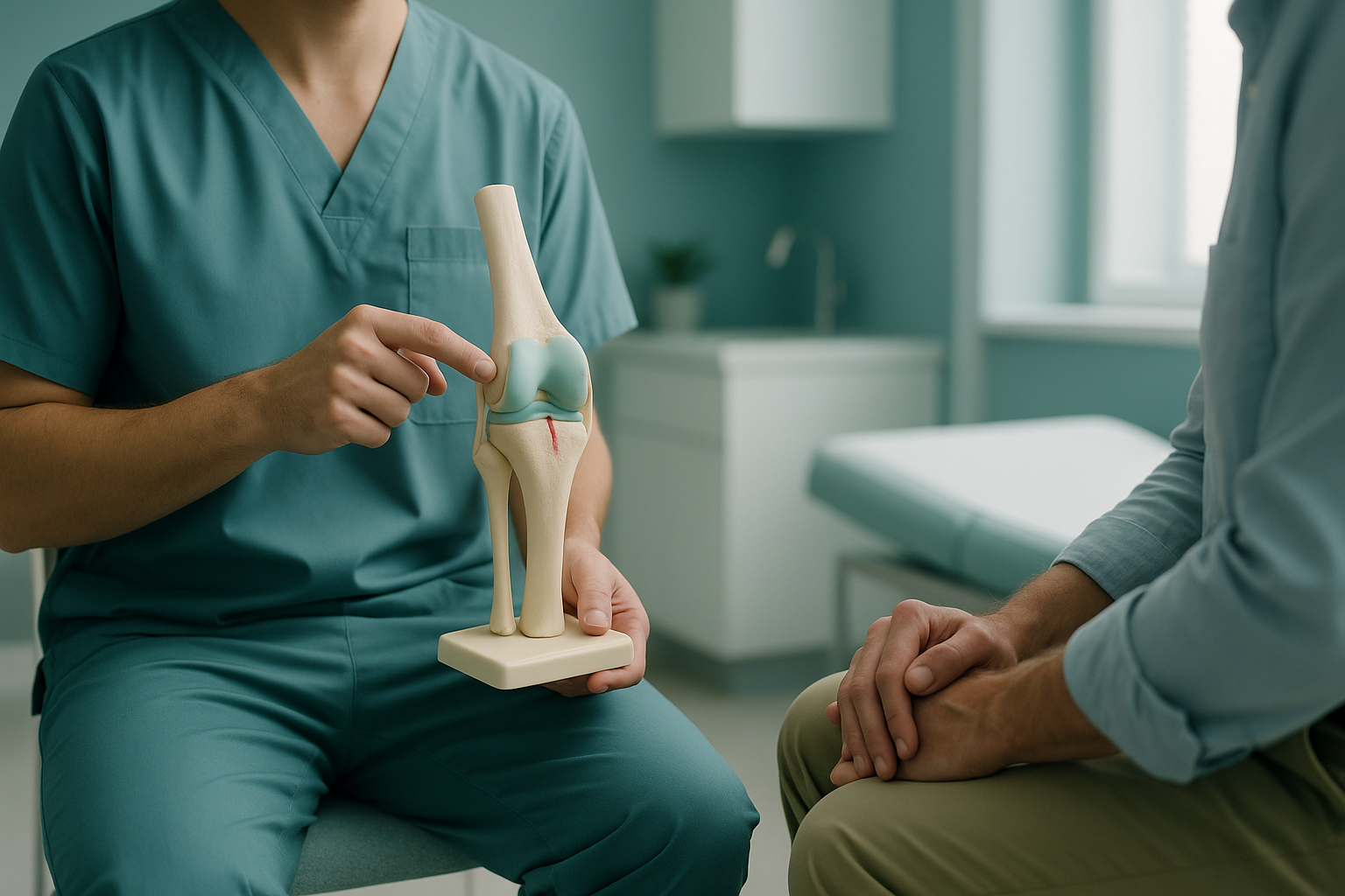
This is the part that standard imaging often misses. An MRI of the knee is taken lying down. In that position, the muscles relax and body weight is taken off the joint. The result is a detailed picture of the tear itself, but no information about the mechanical forces acting on it when you stand and walk.
Your body is a connected chain from your feet to your head. A slight tilt at the hip, or an ankle that rolls inward, changes how force travels through the knee. Pain often shows up in a different place from the real cause. Seeing the whole chain under load reveals patterns that a lying-down scan of a single area cannot.
How does a standing EOS scan help with a meniscus tear?
A standing EOS scan captures your skeleton from head to toe while you carry your full body weight. It shows whether your leg alignment is putting one compartment of the knee under more stress than it should take. This gives your specialist context that lying-down imaging does not provide.
ScanAlign standing imaging uses about 90% less radiation than a standard X-ray, around ten times less. The scan takes just seconds and produces a full-length image of your skeletal alignment under real-life conditions.
Wondering whether alignment might play a part in your knee pain? A free video consultation, which you can have from home before deciding to travel to London, can help you decide if a standing scan is the right next step.
Book a free video consultationAn EOS scan does not replace MRI for confirming the tear itself. It adds the alignment layer. Together, MRI and standing imaging give a complete picture: the state of the soft tissue and the mechanical setting it works in. That combination helps surgeons and physiotherapists plan the most effective approach.
Before you start treatment, or change the one you are on, it is worth getting a detailed diagnosis. Knowing whether the cause sits in your pelvis, your legs or your spine changes what will actually help.
If you are thinking about whether to book a standing knee scan, the free video consultation is a good place to start. A specialist radiographer will review your situation and tell you whether a standing scan would add useful information.
Frequently asked questions about meniscus tears
A meniscus tear is damage to one of the two crescent-shaped cartilage pads that cushion the knee joint. They sit between the thighbone and the shinbone. They help absorb shock and share load across the joint surface.
Common signs include pain along the joint line, swelling, stiffness, and a catching or locking feeling in the knee. A clinical examination and imaging, usually an MRI for soft tissue detail, confirm the diagnosis.
Many degenerative tears improve with a structured rehabilitation programme. The torn tissue itself may not fully heal, but symptoms often settle enough that surgery is not needed. Your clinician will guide you based on how you respond to physiotherapy.
A bucket-handle tear is a large, flap-like tear where part of the meniscus moves out of place into the centre of the joint. It often causes the knee to lock, meaning you cannot fully straighten it. This type usually needs a surgical review.
If your leg is angled so that one side of the knee takes more load than the other, the meniscus on that side is under more stress. Understanding your alignment helps clinicians see why the tear happened and what might stop it from getting worse.
An EOS scan shows your full skeletal alignment under body weight. MRI gives detailed soft tissue images but is taken lying down. So it does not show how load is spread across the knee when you stand. The two scans give information that works together.
Yes. The EOS system uses about 90% less radiation than a standard X-ray, around ten times less. It captures the image in seconds while you stand still.
No. ScanAlign is a private self-pay clinic. You can refer yourself. The process starts with a free video consultation, where a specialist radiographer reviews whether a scan is right for you.
The price for patients travelling from abroad is confirmed during the free video consultation.
If your knee is completely locked and you cannot straighten it, or if you have sudden severe swelling after an injury, seek urgent medical attention from your doctor or an emergency department. A locked knee may need prompt specialist assessment.
Understand your knee, from the ground up
A free video consultation with a specialist radiographer can help you decide whether a standing alignment scan would add useful information to your knee assessment.
Book a free video consultationWritten by Abbas Dhami (Specialist Diagnostic Radiographer)
Sources
- Sihvonen R, Paavola M, Malmivaara A, et al. Arthroscopic partial meniscectomy versus sham surgery for a degenerative meniscal tear. New England Journal of Medicine. 2013;369(26):2515-2524.
- Katz JN, Brophy RH, Chaisson CE, et al. Surgery versus physical therapy for a meniscal tear and osteoarthritis. New England Journal of Medicine. 2013;368(18):1675-1684.
- NHS. Meniscal tear. NHS Conditions. Available at: nhs.uk.
This page is general information and not a diagnosis.
ScanAlign is the trading name of CoreMed Solutions Ltd and operates under The Harley Street Hospital's CQC licence.