
What Is Lumbar Instability, and Why Do Scans Often Miss It?
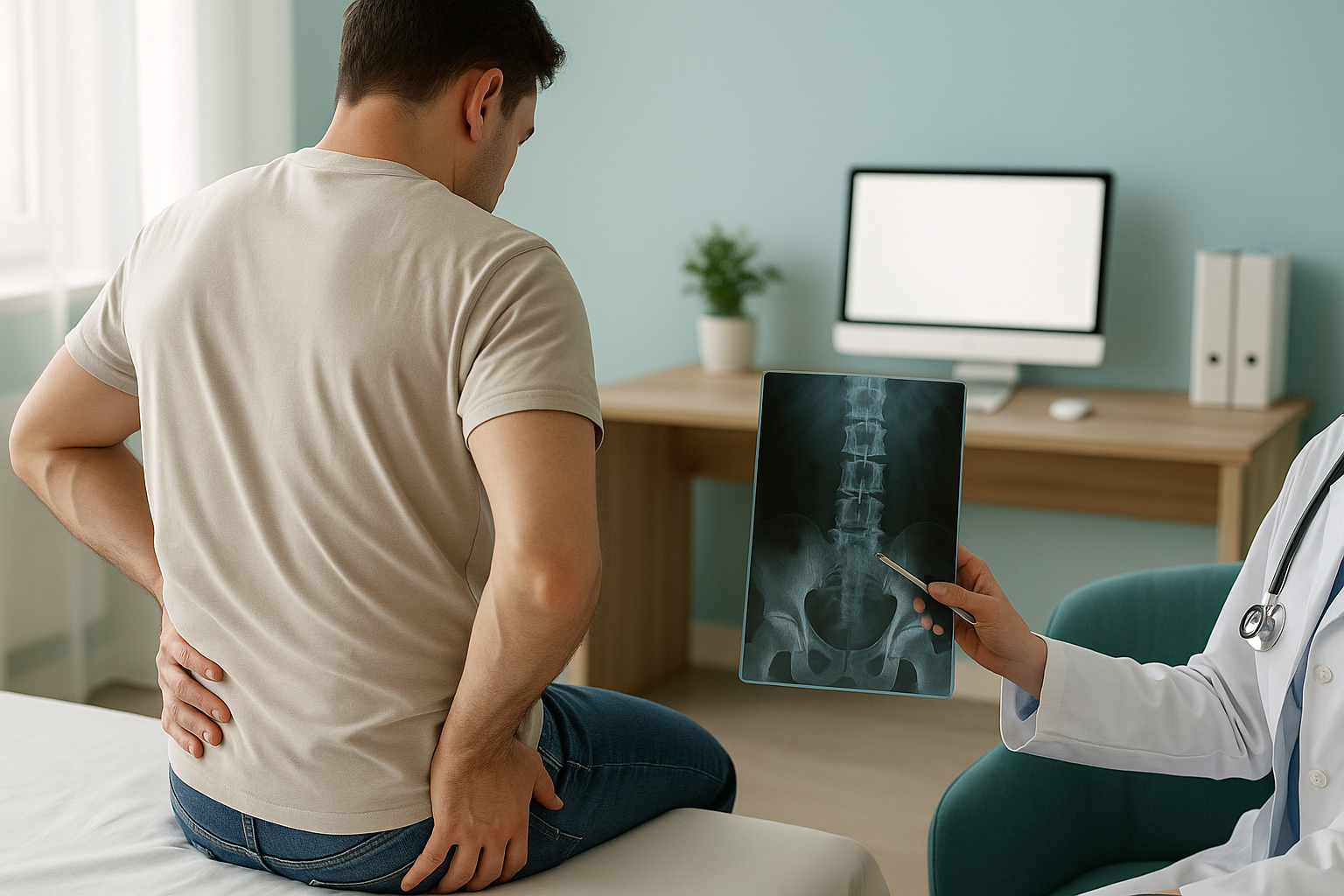
Lumbar instability means one or more vertebrae in your lower back move more than they should when you bend, stand or carry load. It often causes a catching pain that comes and goes. Normal scans are taken lying down. They can look completely normal, because gravity is removed and the muscles are relaxed. This hides the extra movement.
Key takeaways
- Lumbar instability is extra movement between vertebrae. It is not a broken bone or a bulging disc.
- It often causes catching, giving-way or sharp pain with certain movements.
- MRI and CT scans done lying down can miss it, because gravity and body weight are removed.
- Flexion-extension X-rays are the established way to check for segmental movement.
- A standing, weight-bearing scan gives a useful baseline that shows your spine under real load.
What does lumbar instability feel like?
The main sign of spinal instability is a catching or giving-way feeling in the lower back. It tends to be positional. This means it appears with certain movements and goes away at rest. Many people say it feels as if their back "shifts" or "locks" when they bend forward, twist, or stand up from sitting.
A sharp, sudden catch in the lower back when you change position, especially when bending forward or straightening up.
A feeling that your back cannot support you, often after standing or walking for a while. You might need to lean on something.
The pain often eases once you lie down or find a comfortable position. This is why scans taken at rest can look normal.
Your back muscles may spasm to protect the unstable segment. This can feel like tightness or stiffness around the lower spine.
When to get urgent help. If you develop numbness or weakness in your legs, or any loss of bladder or bowel control, contact your doctor or go to the nearest emergency department straight away. These symptoms need immediate medical attention.
What causes unstable back joints?
Segmental instability happens when the structures that hold a vertebra in place can no longer control movement. This can come from wear and tear over time, an old injury, or changes after surgery. Several factors can play a part.
As discs lose height and stiffness, they support the vertebrae less well. This allows more movement.
The small joints at the back of the spine guide movement. When cartilage wears away, these joints can allow too much sliding.
Operations that remove bone or disc material can change the balance of forces. This sometimes makes a segment less stable.
When a vertebra slips forward over the one below, the segment can become unstable. Learn more about spondylolisthesis.
A related condition, retrolisthesis, is when a vertebra slips backward. Both types of slip can cause instability, especially under load.
Why do standard scans miss lumbar instability?
Standard MRI, CT and plain X-rays are usually taken while you lie flat on a table. In that position, gravity is no longer pushing down through your spine, and your muscles relax. An unstable segment can settle back into a normal-looking position. So the images look fine, even though your back moves too much when you are upright.

This is why lumbar instability is sometimes called a "dynamic" problem. It only shows itself with movement or load. A scan taken lying on your back is a snapshot of your spine in its most relaxed, least revealing position.
The established clinical test for segmental instability is a flexion-extension X-ray. Two separate images are taken: one with you bending fully forward and one leaning fully backward. The difference between the two shows how much each segment moves. It is a well-validated approach. But it only captures those two extremes of movement. It does not show your full alignment from head to feet while you stand in a natural posture.
How does standing imaging help assess spinal instability?
A standing, weight-bearing scan captures your spine exactly as it works in daily life. Gravity pulls through every joint and your muscles actively hold you upright. This matters because an unstable segment may slip or shift under that natural load in ways that disappear when you lie down.
At ScanAlign, weight-bearing standing imaging uses low-dose EOS technology to make a full-length image of your skeleton from head to feet while you stand. The dose is about 90% less radiation than a standard X-ray, around ten times less. This gives a clear baseline of your spinal alignment under load.
It is worth being precise here. A single standing image is not the same as a flexion-extension study. That study is still the standard method for measuring segmental movement between two positions. What standing imaging adds is the bigger picture. It shows your overall alignment, pelvic balance, and how your lower spine relates to your hips and legs. An unstable segment does not exist on its own. If one vertebra moves too much, the rest of your spine compensates. Those compensations are only visible when you are standing and bearing your full weight. Understanding weight-bearing versus lying imaging helps explain why this matters.
If you have been told your scans look normal but your back still catches or gives way, a standing assessment can add information that lying-down imaging cannot give.
Book a free video consultationWhen MRI is the better choice. EOS shows bones, joints and alignment. If your clinician suspects a disc bulge pressing on a nerve, or needs to assess the spinal cord or soft tissues, MRI is usually the better tool. The two types of imaging answer different questions and often complement each other.
What can be done about lumbar instability?
Managing lumbar instability depends on knowing exactly what is moving and how much. That is why accurate imaging matters. The right images give your clinician the information they need to plan the most suitable next step. ScanAlign is a diagnostic imaging clinic, so we do not provide treatment. We provide the detailed alignment data that helps the right specialist make better decisions for you.
Common approaches your treating clinician may consider include targeted core-stability physiotherapy to retrain the muscles that support the unstable segment, changes to your activities, or, in more serious cases, a surgical opinion. Clear imaging of your alignment under load helps guide that decision.
If your instability is linked to a vertebral slip, you can read more about spondylolisthesis or retrolisthesis to understand the different directions a vertebra can move.
Before you start a treatment, or change the one you are on, it is worth getting a detailed diagnosis first. Knowing whether the cause sits in your pelvis, your legs or your spine changes what will actually help.
Frequently asked questions about lumbar instability
Lumbar instability is extra movement between two or more vertebrae in the lower back. The structures that normally hold the vertebrae in place, including discs, ligaments and small facet joints, no longer control motion properly. This can cause catching pain, a feeling of giving way, or muscle spasm.
MRI is taken lying down. This removes gravity and lets your muscles relax. An unstable segment can settle back into a normal-looking position on the scan. Instability is a movement problem, so it may not show up when the spine is still and not carrying load.
A flexion-extension X-ray means taking two images of your spine: one bending fully forward and one leaning fully back. Comparing the two shows how much each segment moves. It is the standard clinical method for assessing segmental instability.
No. A standing EOS scan captures your full alignment from head to feet in a natural standing position, under real body weight. It does not measure segmental movement between two extreme positions the way flexion-extension views do. The two complement each other. EOS gives the overall alignment picture, while flexion-extension views measure the movement of specific segments.
EOS uses about 90% less radiation than a standard X-ray, around ten times less. This makes it suitable for full-body imaging without the high dose linked with conventional radiography or CT.
No. ScanAlign is a private, self-pay clinic. You can refer yourself. The process starts with a free video consultation to make sure a standing scan is right for your situation.
It can. As discs keep losing height or facet joints wear further, the amount of movement may increase. Early and accurate imaging helps track changes and supports better clinical decisions about management.
A slipped (or herniated) disc is when the soft inner material of a disc pushes outward and may press on a nerve. Instability is about extra movement of the vertebra itself. You can have one without the other, or both together. They need different types of imaging to assess properly.
The price for patients travelling from abroad is confirmed during your free video consultation, which you can arrange on the book a weight-bearing spine scan page.
A specialist radiographer reviews your images, and a report is written setting out your alignment findings. You can share this report with your treating clinician, physiotherapist or surgeon to help guide the next steps in your care.
Normal scans but your back still catches?
A standing, weight-bearing scan can reveal alignment problems that lying-down imaging misses. Start with a free video consultation.
Book a free video consultationWritten by Abbas Dhami (Specialist Diagnostic Radiographer)
Sources
- Leone A, Guglielmi G, Cassar-Pullicino VN, Bonomo L. "Lumbar intervertebral instability: a review." Radiology, 2007; 245(1):62-77.
- Defined criteria for segmental instability on flexion-extension radiographs. White AA, Panjabi MM. Clinical Biomechanics of the Spine, 2nd ed. Philadelphia: Lippincott, 1990.
- NHS guidance on low back pain assessment and imaging. NICE Clinical Guideline NG59, 2016 (updated 2020).
This page is general information and not a diagnosis.
ScanAlign is the trading name of CoreMed Solutions Ltd and operates under The Harley Street Hospital's CQC licence.