
What Is an LCL Injury, and How Is It Different From an MCL Tear?
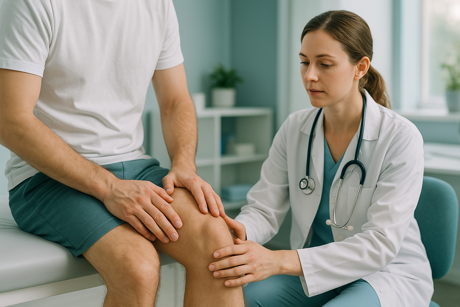
An LCL injury is damage to the lateral collateral ligament. This is the tough band on the outer side of your knee. It usually happens when a force pushes the knee inward. An MCL injury affects the inner side. LCL tears are less common than MCL tears, but they are more likely to involve nearby structures such as the posterolateral corner. This makes an accurate diagnosis important.
Key points
- The LCL keeps the outer side of the knee stable and stops it from bowing outward.
- Injuries range from a mild stretch (grade 1) to a complete tear (grade 3).
- LCL tears often happen together with damage to the posterolateral corner or a PCL strain.
- Standing, weight-bearing imaging can show alignment problems that lying-down scans miss.
- If you feel numbness, weakness or loss of control of your leg, get urgent medical help.
What exactly is the lateral collateral ligament?
The lateral collateral ligament (LCL) is a cord-like band. It runs down the outer side of your knee and connects the thighbone to the smaller bone of the lower leg. Its main job is to stop the knee from bowing outward when you stand, walk or change direction.
The LCL works as part of a group of structures called the posterolateral corner. This group includes other ligaments, tendons and a small muscle. Together they keep the outside and back of the knee stable. When the LCL is damaged, one or more of these nearby structures may be injured too. This is why an LCL tear can feel more complex than a simple sprain.
Grades of LCL injury
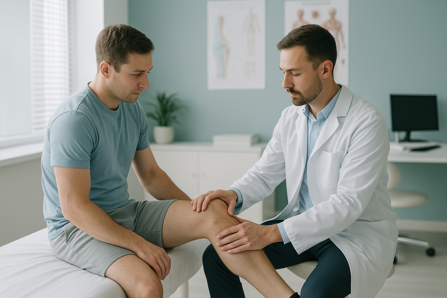
- Grade 1: The ligament is stretched but not torn. The outside of the knee may feel tender, but the joint stays stable.
- Grade 2: A partial tear. The outer knee feels loose during certain movements, and the swelling is more noticeable.
- Grade 3: A complete tear. The knee may give way when you stand or twist. Other structures in the posterolateral corner are often affected.
What does an LCL tear feel like?
An LCL tear typically causes a sharp pain on the outer side of the knee. It is often felt at the moment of injury. Over the next few hours the area may swell, stiffen and feel unstable.
Tenderness or a burning feeling along the outside of the knee. It is often worse when you press on the area.
The knee may buckle outward when you walk, especially on uneven ground or stairs.
Mild to moderate swelling on the outer knee. Sometimes bruising appears a day or two later.
Locking or stiffness that makes it hard to straighten the knee fully, especially after rest.
When to get urgent help. If you notice numbness down your lower leg, foot drop or sudden weakness, the peroneal nerve near the LCL may be involved. Contact your doctor or go to an emergency department without delay.
What causes an LCL injury?
An LCL injury most often happens when a strong force pushes the knee inward. This stretches the outer ligament beyond its limit. It can also develop slowly through repeated stress on a knee that is already poorly aligned.
A blow to the inside of the knee during rugby, football or martial arts.
A sudden change of direction with the foot planted, common in tennis or skiing.
Long-standing varus alignment (bow-legged posture) puts extra strain on the outer ligament over time.
The body works as a connected chain from the feet to the head. So an alignment problem in the hip or ankle can shift extra load onto the outer knee. Over months or years, this added stress makes the LCL more vulnerable, even without one obvious injury.
How does an LCL injury differ from an MCL tear?
The LCL sits on the outer side of the knee, while an MCL injury affects the inner side. Both are collateral ligament injuries. But they differ in how they happen, how common they are and what else tends to get hurt with them.
Location: Outside of the knee.
Typical cause: A force pushing the knee inward, or a twisting injury.
How common: Less common than MCL injuries.
Related damage: Often involves the posterolateral corner, the PCL strain structures, or the peroneal nerve.
Healing: Grade 3 tears more often need a surgical assessment, because the LCL has a poorer blood supply than the MCL.
Location: Inside of the knee.
Typical cause: A blow to the outer knee pushing it inward, common in football.
How common: The most commonly injured knee ligament.
Related damage: May occur with an ACL tear in more severe cases.
Healing: The MCL has a good blood supply, so many tears heal with bracing and rehabilitation alone.
Why does a standing scan matter for an LCL injury?
A standing, weight-bearing scan shows how your knee alignment behaves under real-life load. When you lie down for a standard X-ray or MRI, your muscles relax and gravity no longer pulls on the joint. This can hide the outward bowing (varus thrust) that stresses the LCL every time you walk.
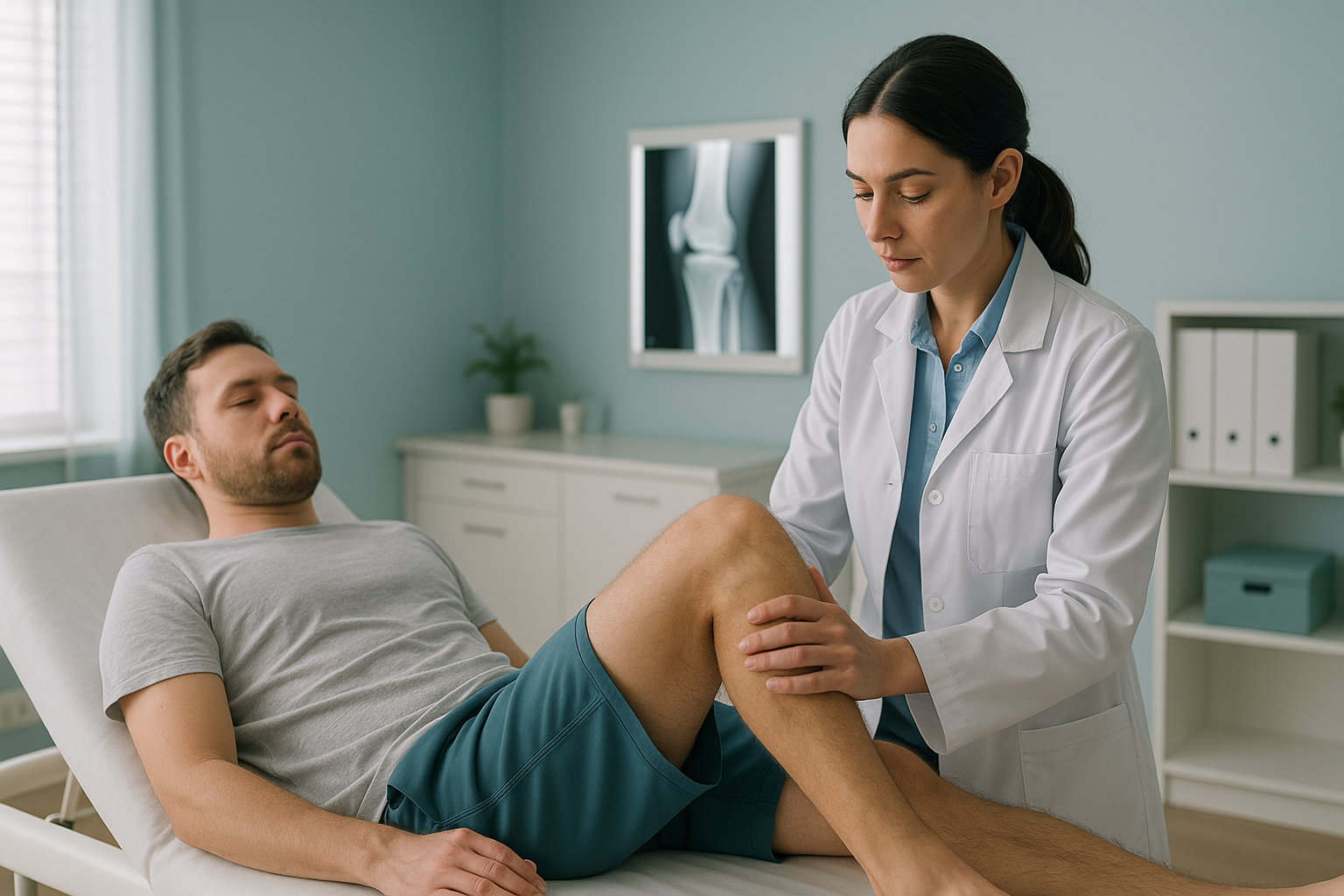
ScanAlign standing EOS imaging captures your full leg alignment from hip to ankle in a single upright image. It uses about 90% less radiation than a standard X-ray, around ten times less. This gives your clinician the information they need to see whether varus alignment or a gait thrust is overloading your LCL.
Want to find out if your knee alignment is adding to your outer knee pain?
Book a free video consultationWhat generally helps after an LCL injury?
Treatment depends on the grade of the tear and whether other structures are involved. ScanAlign is a diagnostic imaging clinic, so we do not provide treatment. But knowing what options usually exist may help you ask the right questions at your next appointment.
- Rest, ice and protection: Mild sprains often settle with changes to activity, ice and a supportive brace.
- Physiotherapy: Guided rehabilitation strengthens the muscles around the knee and restores confidence in the joint.
- Bracing: A hinged brace can limit sideways movement while the ligament heals.
- Surgical assessment: Complete tears, especially those involving the posterolateral corner, may be referred for a surgical opinion.
- Alignment assessment: Understanding the full picture, including hip, knee and ankle alignment, helps clinicians plan rehabilitation or surgery more accurately.
Before you start treatment, or change the one you are on, it is worth getting a detailed diagnosis. Knowing whether the cause sits in the pelvis, the legs or the spine changes what will actually help.
When MRI is the better tool. EOS shows bones, joints and alignment. For soft tissue detail, such as the ligament fibres themselves, the cartilage around them or nerve involvement, MRI is usually the better choice. Many people benefit from both: an MRI for the soft tissue and a standing EOS scan for the alignment picture.
Frequently asked questions about LCL injuries
Grade 1 and some grade 2 LCL injuries can heal with rest, bracing and physiotherapy. Complete (grade 3) tears, especially those involving the posterolateral corner, are more likely to need a specialist assessment and may need surgery.
LCL pain is usually felt along the outer edge of the knee, roughly level with or just above the joint line. A clinician may do a varus stress test, gently pushing the knee inward while the leg is straight, to check for looseness on the outer side.
The posterolateral corner is a group of ligaments, tendons and a small muscle at the back and outside of the knee. These structures work with the LCL to control rotation and sideways movement. When the LCL tears completely, these structures are often injured too.
Not necessarily "worse", but LCL tears tend to be more complex. The LCL has a poorer blood supply, so it heals less readily than the MCL. LCL injuries also often involve nearby structures, which can make recovery longer.
Yes. Bow-legged (varus) alignment puts extra stress on the outer knee with every step. Over time this can stretch or weaken the LCL. This makes it more vulnerable to injury, even from fairly minor forces.
A lying-down scan removes the effect of gravity and body weight, so it can underestimate how much the knee bows outward. A standing EOS scan captures your leg alignment under real load. It shows varus thrust and other patterns that matter for treatment planning.
Yes. EOS uses about 90% less radiation than a standard X-ray, around ten times less. The scan takes just a few seconds and there is no enclosed tunnel.
No. ScanAlign is a private, self-pay clinic. You can book a standing knee scan directly without a referral.
The price for patients travelling from abroad is confirmed during the free video consultation. You can read more on our FAQs page.
Yes. An unstable outer knee can cause a lateral thrust, a visible outward lurch, during walking. This changed walking pattern loads other joints differently. Over time it can lead to compensatory pain in the hip, ankle or lower back.
See your full leg alignment, standing
In a free video consultation you can talk about your outer knee pain and find out whether a standing EOS scan in London could help.
Book a free video consultationSources
- LaPrade RF, et al. "The posterolateral attachments of the knee." The American Journal of Sports Medicine, 2003; 31(6): 854-860.
- NHS. "Sprains and strains." nhs.uk, 2023.
- Illés T, Somoskeöy S. "The EOS imaging system and its uses in daily orthopaedic practice." International Orthopaedics, 2012; 36(7): 1325-1331.
Written by Abbas Dhami (Specialist Diagnostic Radiographer)
This page is general information and not a diagnosis.
ScanAlign is the trading name of CoreMed Solutions Ltd and operates under The Harley Street Hospital's CQC licence.