

How Does Leg Alignment Cause Knee Osteoarthritis?
When your legs are not straight, your body weight is pushed unevenly through the knee joint. The overloaded side wears its cartilage faster. This narrows the joint space and drives knee osteoarthritis. A full-leg standing X-ray can reveal this imbalance, so treatment targets the real cause, not just the pain.
Key points
- Knee osteoarthritis is the most common form of arthritis. It is caused by cartilage wearing down inside the joint.
- Leg alignment decides how your weight is spread across the knee. Even a small tilt shifts load to one side.
- Alignment problems only show up reliably when you are standing and bearing weight, not lying down.
- Understanding your alignment helps clinicians choose the right approach, from bracing to surgery.
- EOS standing imaging captures the full mechanical axis of both legs in a single low-dose scan.
What is knee osteoarthritis?
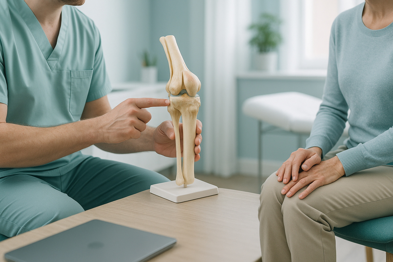
Knee osteoarthritis is a gradual wearing down of the cartilage that cushions the ends of the bones inside your knee joint. Cartilage is a smooth, tough tissue that lets the joint glide freely. When it thins or breaks down, the bones underneath come closer together. The joint space narrows, and movement becomes painful and stiff.
It is the most common type of arthritis in the UK and tends to develop over years. Most people first notice it after middle age. Younger adults can develop it too, especially after an injury or if their legs are not well aligned.
What are the common symptoms of knee osteoarthritis?
Symptoms usually build slowly. You might notice one or two at first, then more over time.
Your knee feels tight when you first stand up, especially in the morning or after sitting for a while.
Aching or sharp pain during weight-bearing activities, often on one side of the knee.
The joint may look puffy or feel warm to the touch after activity.
A rough, crunching feeling when you bend or straighten the knee, caused by uneven bone surfaces.
If you notice sudden weakness, locking, or giving way of the knee, see your doctor, or go to an emergency department if it follows an injury. These symptoms need prompt assessment.
How does leg alignment make knee arthritis worse?
Your lower limb alignment controls where your body weight lands inside the knee. In a well-aligned leg, the mechanical axis (an imaginary line from the hip centre to the ankle centre) runs roughly through the middle of the knee. This spreads the load evenly across both sides of the joint.
When that line shifts, one compartment of the knee takes far more than its fair share of force. Over months and years, the overloaded cartilage wears faster than the body can repair it.
The mechanical axis falls to the inner side. The inner (medial) compartment is overloaded. This is the most common pattern in knee osteoarthritis.
The mechanical axis falls to the outer side. The outer (lateral) compartment takes the extra force.
Subtle rotation in the shin or thigh bone can create shearing forces that speed up wear, even when alignment looks straight from the front.
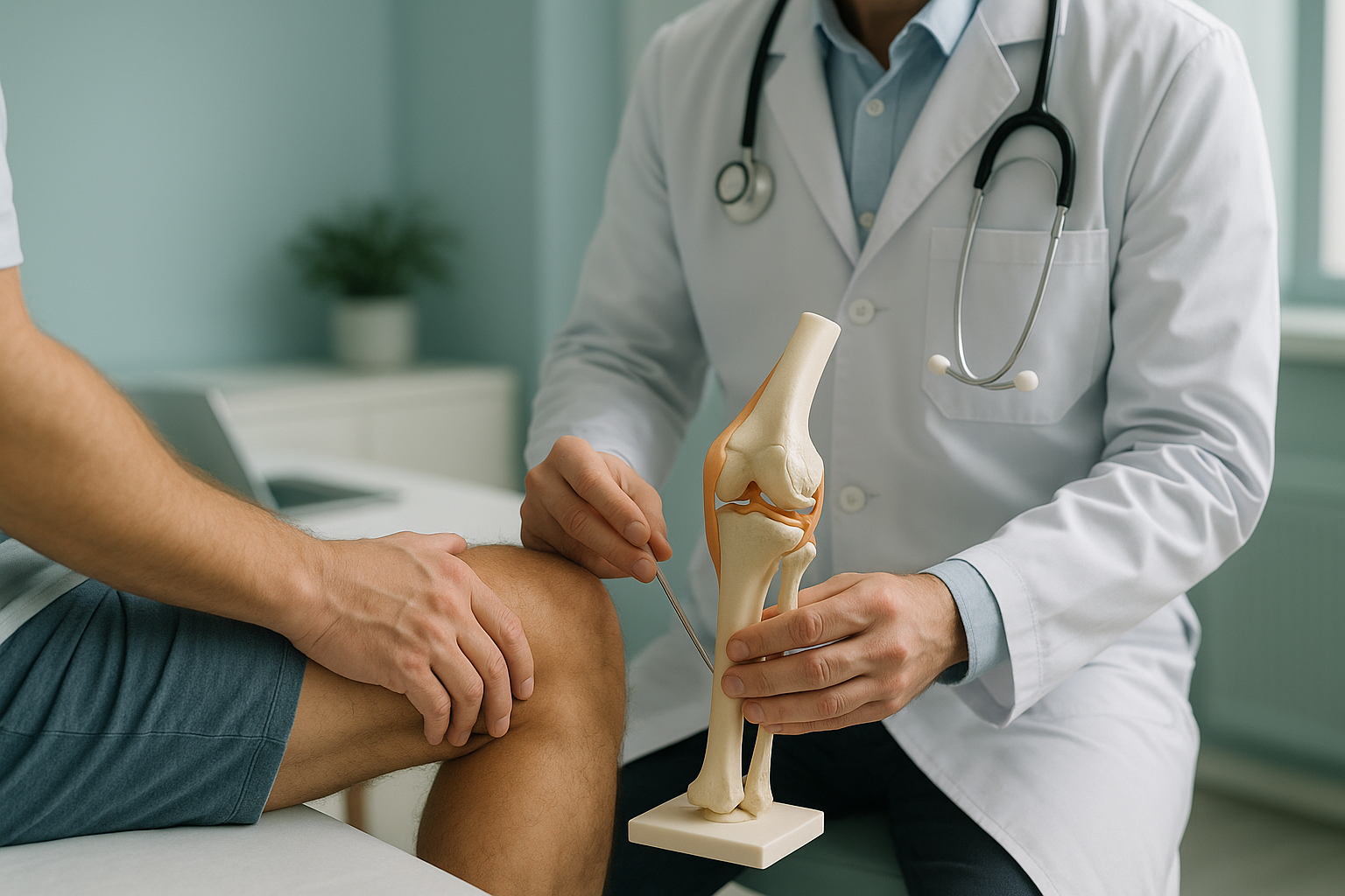
This is not just a theory. Research consistently shows that even small shifts in alignment speed up cartilage loss and joint space narrowing. It also works the other way. Arthritis itself can worsen alignment, as worn cartilage makes the joint tilt further. The two problems feed each other.
That is why it matters to understand whether your knee pain is driven by knee alignment or osteoarthritis, or both together. It helps you choose the right path forward.
Why does a standing scan show alignment problems that lying-down imaging misses?
Standard X-rays, CT and MRI are usually taken with you lying flat. In that position, gravity is not pressing down through the joint. Muscles relax. The joint can settle into a position that looks more aligned than it really is. Any tilt, rotation or uneven loading disappears.
When you stand, your full body weight compresses the joint. The true gap between the bones becomes visible, and any shift in the mechanical axis is revealed. This is the position your knee actually works in, so it is the position that matters for planning treatment.
You stand naturally in the EOS scanner. It captures both legs from hip to ankle in a single image, showing the full mechanical axis under real load. Cartilage loss, joint space narrowing and malalignment are all visible as they truly are. The scan uses about 90% less radiation than a standard X-ray, around ten times less.
Lying-down X-rays can show bone spurs and rough joint surfaces. But they cannot show how your weight is spread through the joint. The alignment may look acceptable on the table but shift a lot when you stand. CT and MRI add soft-tissue detail, but they share the same limit: they are not weight-bearing.
ScanAlign EOS imaging captures the full picture in one standing exposure. The measurements give your surgeon or physiotherapist the data they need to decide on bracing, exercise, realignment surgery or joint replacement, and to plan it accurately.
When MRI is the better choice: EOS shows bones, joints and alignment clearly. If your clinician suspects soft-tissue damage, meniscus tears, ligament injuries or nerve involvement, MRI is usually the better tool. The two scans answer different questions and often work well together.
What helps manage knee osteoarthritis when alignment is involved?
Treatment decisions depend on how severe the arthritis is and how much malalignment there is. ScanAlign does not provide treatment. But the alignment data from a standing scan helps your clinician choose from several well-established approaches.
- Have knee arthritis that seems worse on one side of the joint
- Are considering knee replacement and want planning data before the operation
- Have been told your legs look bowed or knock-kneed
- Want to understand whether alignment is contributing to your symptoms
- Have had a standard X-ray but still do not have a clear explanation
- Suspect a meniscus tear, ligament injury or soft-tissue problem (MRI is more appropriate)
- Have sudden locking, giving way, or significant swelling after an injury (see your doctor or go to an emergency department)
- Need blood tests to rule out inflammatory arthritis such as rheumatoid arthritis
Before you start a treatment, or change the one you are on, it is worth getting a detailed diagnosis. Knowing whether the cause sits in the pelvis, the legs or the spine changes what will actually help.
Not sure whether a standing alignment scan is right for your knee? Our specialist radiographer can talk through your situation in a short video call from wherever you are, at no cost and with no obligation.
Book a free video consultationFrequently asked questions about knee osteoarthritis and alignment
Yes. When the mechanical axis of the leg is shifted, one side of the knee carries more load than the other. Over time, the overloaded cartilage wears faster. Research shows that malalignment is one of the strongest risk factors for the progression of knee osteoarthritis.
Varus means the knee angles inward, creating a bow-legged look. It overloads the inner compartment. Valgus means the knee angles outward, creating a knock-kneed look, and overloads the outer compartment. Both patterns speed up cartilage wear on the affected side.
A normal X-ray taken lying down removes the effect of gravity. The joint space can look wider, and alignment can look more normal than it really is. A standing X-ray shows how the joint behaves under your actual body weight. This gives a much more accurate picture of load distribution and cartilage loss.
EOS uses about 90% less radiation than a standard X-ray, around ten times less. It captures a full-length image of both legs in a single low-dose exposure. It is one of the lowest-radiation options available for this type of imaging.
No. ScanAlign accepts self-referrals. You can book directly without seeing your doctor first. If you like, you are welcome to bring a referral letter, but it is not required.
For patients travelling from abroad, the price is confirmed during the free video consultation, which you can arrange on the book a standing knee alignment scan page. ScanAlign is a private self-pay clinic.
In some cases, targeted exercises, orthotics or unloader braces can reduce the load on the affected compartment. The right approach depends on how severe both the alignment shift and the arthritis are. A standing alignment scan helps your clinician decide which option is most appropriate for you.
The scan provides detailed alignment and joint-space data that surgeons use when planning a knee replacement. It does not replace a clinical consultation. But it gives your surgeon the measurements they need to assess whether replacement is appropriate and to plan it precisely.
Osteoarthritis is a mechanical wear-and-tear condition, often made worse by alignment. Inflammatory arthritis (such as rheumatoid arthritis) is driven by the immune system. It usually needs blood tests and specialist rheumatology assessment. An EOS scan is designed for mechanical and alignment assessment, not for diagnosing inflammatory conditions.
Yes. The EOS scanner captures both legs from hip to ankle in a single standing image. This also lets your clinician compare one side with the other, which helps with planning if one knee is more affected.
See how your alignment affects your knee
A short video call with our specialist radiographer can help you understand whether a standing alignment scan in London is the right next step for your knee.
Book a free video consultationSources
- Sharma L, et al. "The role of knee alignment in disease progression and functional decline in knee osteoarthritis." JAMA, 2001; 286(2): 188-195.
- National Institute for Health and Care Excellence (NICE). "Osteoarthritis: care and management." Clinical guideline CG177, updated 2022.
- Illés T, Somoskeöy S. "The EOS imaging system and its uses in daily orthopaedic practice." International Orthopaedics, 2012; 36(7): 1405-1411.
Written by Abbas Dhami (Specialist Diagnostic Radiographer)
This page is general information and not a diagnosis.
ScanAlign is the trading name of CoreMed Solutions Ltd and operates under The Harley Street Hospital's CQC licence.