
How Does Imaging Support Rheumatology Assessment?
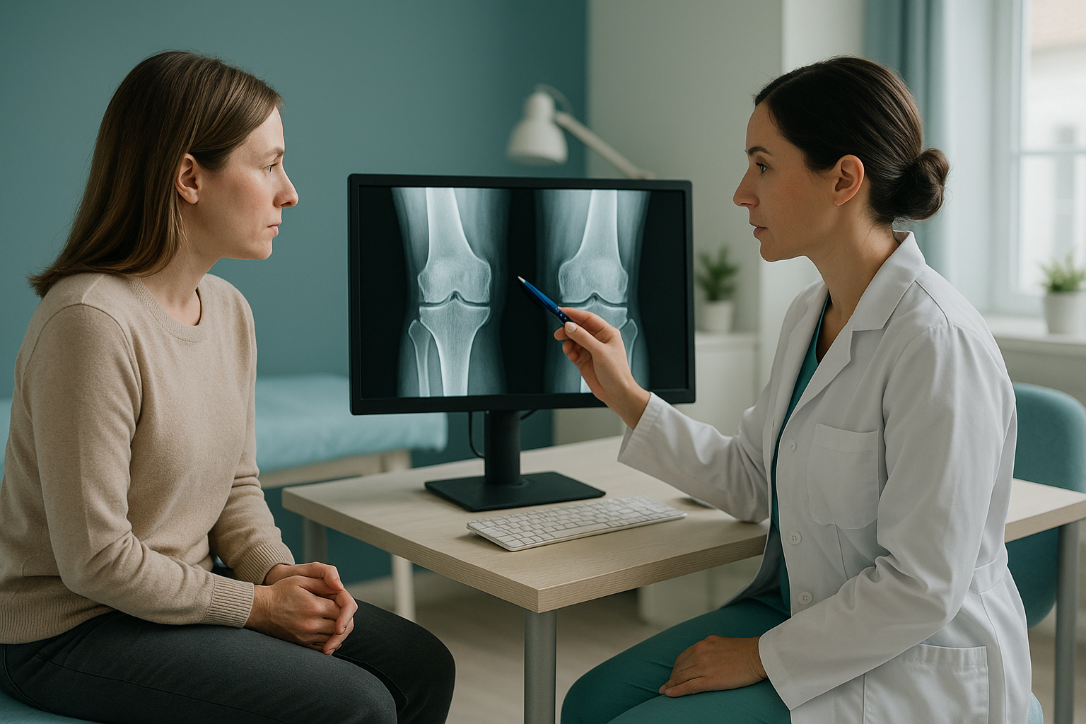
Rheumatology assessment uses different imaging tools for different questions. MRI detects active inflammation, such as sacroiliitis and bone marrow oedema. Standing whole-body imaging adds structural context. It captures sagittal balance, changes in spinal mobility and the mechanical effects of disease under weight-bearing conditions. Lying-down scans cannot show these.
Key takeaways
- MRI remains the standard for detecting active inflammation in axial spondyloarthritis. EOS imaging does not replace it.
- Standing EOS imaging shows structural change, whole-spine alignment and sagittal parameters under real weight-bearing load.
- Low-dose repeat imaging lets you monitor how spinal disease progresses over time, with about 90% less radiation than a standard X-ray.
- Reports include full sagittal measurements and can be shared directly with the referring rheumatologist.
- No referral from a family doctor is needed. You can register to refer patients directly.
What is the role of MRI in rheumatology imaging?
MRI is the main tool for detecting active inflammation. In axial spondyloarthritis, MRI of the sacroiliac joints can identify bone marrow oedema and early sacroiliitis before structural damage shows on ordinary X-rays. It is also essential for assessing soft tissue, discs and nerves.
Clear facts on what EOS does and does not do. EOS imaging shows bones, joints and structural alignment. It does not detect active inflammation, bone marrow oedema or soft-tissue disease. For those questions, MRI is usually the better tool. We are open about this because the two methods answer different clinical questions, and both may be needed.
Standing imaging adds value in the structural questions MRI cannot easily answer. These include overall spinal alignment, load-bearing posture and how disease affects the whole kinetic chain from pelvis to skull.
Why does standing imaging matter for spinal disease?
Standing imaging captures the spine under the load of gravity, which is how your patient actually lives. Lying-down MRI and CT relax the paraspinal muscles and for a while flatten compensatory curves. So supine imaging can underestimate the true extent of sagittal imbalance, kyphosis progression or pelvic compensation.
What you see: True weight-bearing alignment from head to feet in a single calibrated image. Full sagittal parameters, including pelvic incidence, sacral slope, lumbar lordosis and thoracic kyphosis. Compensatory mechanisms such as pelvic retroversion, knee flexion and cervical hyperextension are visible because the patient is standing naturally.
What you see: Excellent soft-tissue contrast, inflammation and disc detail. However, gravity is removed. Compensatory postures relax, kyphotic deformity may look less severe, and the link between the pelvis and spine is not under load. Overall alignment from feet to skull is not captured in a single acquisition.
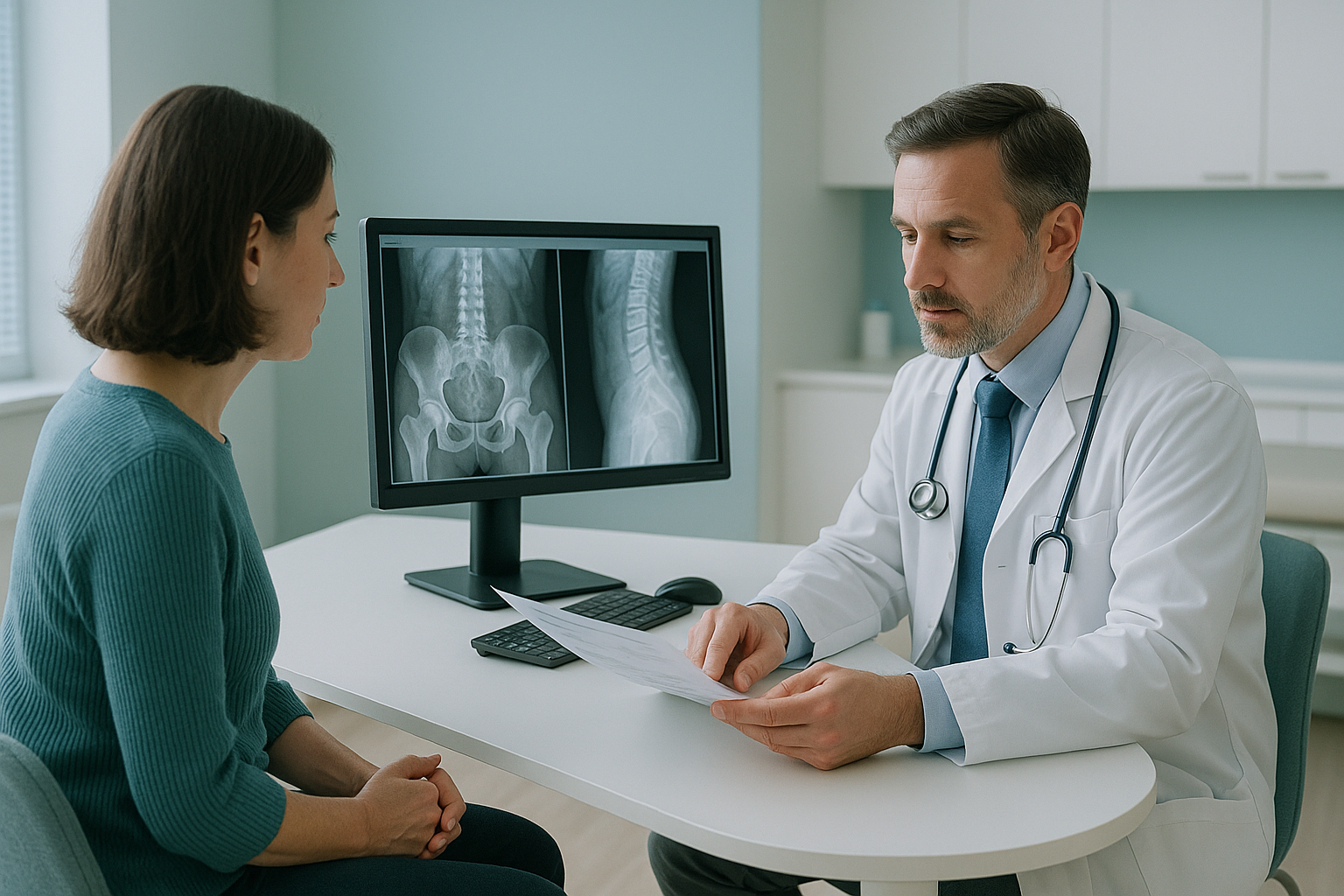
The EOS system uses slot-scanning biplanar technology with about 90% less radiation than a standard X-ray, around ten times less. This makes it practical for repeat imaging to monitor structural change over months or years without concern about cumulative dose.
How can rheumatologists use standing alignment data?
Standing whole-body imaging gives clinical data that adds to your MRI and blood tests. Below are the most common rheumatology referral cases we see.
Record sagittal parameters at diagnosis to set a structural reference point before starting biologics or other therapies.
Repeat low-dose scans track kyphosis, loss of lumbar lordosis and sagittal vertical axis shift over time. This supports clinical correlation with disease activity scores.
When a patient with ankylosing spondylitis is being considered for corrective osteotomy, the surgeon needs standing full-spine imaging with pelvic parameters.
Weight-bearing sacroiliac imaging shows joint space changes, ankylosis and structural fusion. It adds to MRI findings of active inflammation.
How does a rheumatologist refer a patient for standing imaging?
The referral process is simple. No letter from a family doctor is needed. Rheumatologists can refer directly.
Register with ScanAlign
Complete a short one-time registration so we can link reports to your practice. You can register to refer patients online.
Send a referral
Send your clinical question and the patient's details. State the regions of interest and any sagittal parameters you need.
Patient comes to London for imaging
The scan takes place at 19 Harley Street, London. The whole-body standing acquisition takes only a few seconds, with about 90% less radiation than a standard X-ray.
Shared reporting
The report, including full sagittal parameter measurements, is sent directly to you. You can then compare it with your disease activity assessments and MRI findings.
Which rheumatology patients benefit from standing imaging?
Standing whole-body imaging is most useful when structural alignment or mechanical change is part of the clinical question. It does not replace MRI when inflammation is the main concern.
Before a patient starts treatment, or changes the one they are on, a detailed diagnosis is worth having. Knowing whether the cause sits in the pelvis, the legs or the spine changes what will actually help.
- Established axial spondyloarthritis, tracking structural change
- Planning before spinal osteotomy
- Monitoring kyphosis progression or loss of lordosis
- Assessing compensatory changes in the pelvis or legs
- Repeat imaging where cumulative radiation dose matters
- Suspected early sacroiliitis with no structural changes yet
- Detecting active bone marrow oedema or enthesitis
- Assessing soft-tissue inflammation or nerve compression
- Diagnosis of inflammatory arthritis where imaging shows early inflammation only
Would you like to discuss whether standing imaging would add value for a particular patient? We are happy to talk through the clinical question first.
Refer a patientFrequently asked questions about rheumatology imaging referrals
No. EOS imaging shows bone structure, joint spaces and alignment. It does not detect active inflammation, bone marrow oedema or soft-tissue swelling. MRI is the right tool for those findings.
EOS uses about 90% less radiation than a standard X-ray, around ten times less. This matters most for repeat imaging in chronic conditions where cumulative dose is a concern.
No. Rheumatologists and other specialists can refer directly. Patients can also refer themselves. There is no need for a letter from a family doctor.
The report includes pelvic incidence, sacral slope, pelvic tilt, lumbar lordosis, thoracic kyphosis, sagittal vertical axis and other relevant measurements. These are calibrated from a weight-bearing, full-body acquisition.
Yes. Because the dose is so low, scans can be repeated at intervals to monitor structural changes such as increasing kyphosis, loss of lordosis or progressive sacroiliac fusion, without significant cumulative radiation exposure.
It is useful for structural assessment of the sacroiliac joints, including joint space narrowing, erosions and ankylosis. However, it does not show active inflammation. MRI remains the standard for detecting early sacroiliitis.
The standing acquisition itself takes only a few seconds. The whole appointment, including positioning and any extra views, is usually short and simple for the patient.
You can complete a short online registration. Once registered, you can send referrals and receive reports directly.
ScanAlign, low dose standing imaging, is located at 19 Harley Street, London. It operates under The Harley Street Hospital's CQC licence.
ScanAlign is a private, self-pay clinic. The price for patients travelling from abroad is confirmed during the free video consultation.
Ready to refer a patient for standing imaging?
Register once, then send referrals directly. Reports include full sagittal parameters and are shared with you for clinical correlation.
Written by Abbas Dhami (Specialist Diagnostic Radiographer)
Sources
- Sieper J, Poddubnyy D. Axial spondyloarthritis. The Lancet. 2017;390(10089):73-84.
- National Institute for Health and Care Excellence (NICE). Spondyloarthritis in over 16s: diagnosis and management (NG65). 2017.
- Dubousset J, Charpak G, Dorion I, et al. A new 2D and 3D imaging approach to musculoskeletal physiology and pathology with low-dose radiation in standing position: the EOS system. Bulletin de l'Académie Nationale de Médecine. 2005;189(2):287-300.
This page is general information and not a diagnosis.
ScanAlign is the trading name of CoreMed Solutions Ltd and operates under The Harley Street Hospital's CQC licence.