

What Are the Symptoms and Stages of Hip Osteoarthritis?
Hip osteoarthritis is the slow wearing of the cartilage inside your hip joint. The first signs are usually a deep ache in the groin or the front of the thigh, stiffness first thing in the morning, and trouble with movements like putting on shoes. It moves through mild, moderate and severe stages. A standing X-ray can show joint changes that a lying-down scan may miss.
Key points
- Groin or thigh ache, morning stiffness and less range of movement are the main early signs of hip arthritis.
- The condition moves through mild, moderate and severe stages. Each stage affects daily life in a different way.
- Lying-down imaging relaxes the joint and can understate how much space has been lost. Standing imaging shows your hip under real load.
- ScanAlign is a diagnostic clinic. It provides the imaging your specialist needs, not treatment itself.
- If you have sudden severe hip pain, leg weakness, or loss of bladder or bowel control, get urgent medical help from your doctor or the nearest emergency department.
Hip osteoarthritis is the most common form of arthritis in the hip. The smooth cartilage that cushions the ball (femoral head) and socket (acetabulum) wears down over time. Bone may rub on bone, small bony growths called osteophytes can form, and the joint space narrows.
This is not the same as a sudden injury. It develops slowly, often over years. Many people live with it for a long time before looking for an answer. Knowing the symptoms and stages helps you have a clearer talk with your clinician. Clearer imaging helps your clinician plan the right next step.
What does arthritis in the hip feel like?
Hip arthritis symptoms usually start with a dull, deep ache, not a sharp pain. It is most often felt in the groin, the front of the thigh or the buttock. It can also be felt in the knee. Many people notice it after sitting for a long time or when getting out of a car.
A deep, nagging ache in the groin or front of the thigh, often worse after activity.
The joint feels stiff when you wake up or after sitting. It usually eases within 30 minutes of moving.
Trouble turning the leg inwards, crossing your legs, or bending down to tie a shoe.
You may start to limp, lean to one side, or take shorter steps without realising it.
Some people also hear or feel a grinding in the joint. Pain at night, especially when lying on the affected side, is common in later stages. If your hip pain is changing how you walk, it may also be changing the load on your knee, pelvis and lower back. This link between hip pain and alignment matters, because your body adjusts as a whole chain.
What are the stages of hip osteoarthritis?
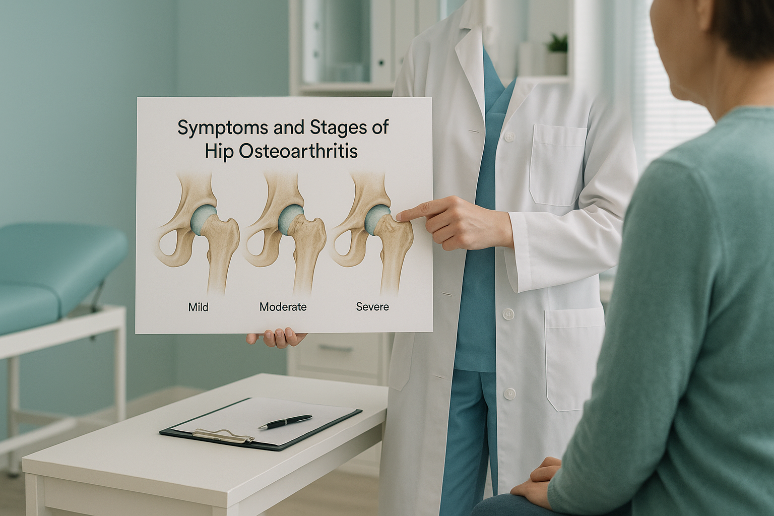
Hip osteoarthritis is usually described in three broad stages: mild, moderate and severe. These are based on how much cartilage is lost, the size of the bony changes, and the effect on daily life.
Mild (early) stage
Cartilage starts to thin, but the joint space is only slightly smaller. You might notice stiffness in the morning or a dull ache after a long walk. Standard X-rays taken lying down may look almost normal at this point. This is because the joint is unloaded, and the gap looks wider than it really is under your body weight.
Moderate stage
The cartilage is clearly thinner. Osteophytes (small bony spurs) start to form around the rim of the joint. Pain comes more often and more predictably, during everyday tasks like climbing stairs or getting up from a chair. You may notice your hip cannot turn as far as it used to.
Severe (advanced) stage
Little or no cartilage is left, and the joint is described as "bone on bone." Pain can be constant, including at rest and at night. How far you can walk drops a lot, and limping is common. This is the stage where a surgeon may discuss hip replacement explained as an option.
What causes hip osteoarthritis?
There is rarely a single cause. Hip osteoarthritis usually develops from a mix of factors over many years.
Cartilage naturally thins as you get older, especially after 50.
A family history of osteoarthritis raises your risk.
Conditions like hip impingement (FAI) or hip dysplasia in adults change how forces pass through the joint.
A fracture or a serious soft-tissue injury can speed up cartilage loss in that hip.
Extra body weight increases the load through the hip with every step. Physically hard jobs or high-impact sports over many years can add to the stress over time. Importantly, a hip joint that is slightly out of alignment may wear unevenly for decades before symptoms appear.
How does a standing X-ray help assess hip arthritis?
A standing X-ray shows your hip joint under your actual body weight, which is exactly how it works in real life. When you lie down for a standard X-ray, CT or MRI, gravity is removed, the muscles relax, and the joint space can look wider than it really is. This means early or moderate narrowing may be understated.
When you stand, your full body weight presses on the hip joint. The image shows the true joint space, any tilt in the pelvis, and how the hip relates to the knee below and the spine above. This gives your clinician a realistic picture of the wear pattern.
Lying flat removes gravity. The femoral head sits differently in the socket, muscles relax, and the pelvis may level out. The image can make the joint gap look bigger than it is when you are on your feet. This may understate the stage of arthritis.
EOS is a low-dose standing imaging system that captures your full skeleton from head to feet in a single scan. It uses about 90% less radiation than a standard X-ray, around ten times less. Because it sees the whole chain, it can show whether a pelvic tilt, a leg-length difference, or spinal alignment is putting extra stress on one hip. ScanAlign standing EOS imaging at 19 Harley Street, London, offers this type of assessment to adults who book themselves.
Thinking of travelling to London and wondering whether a standing scan could give you a clearer picture? Talk to our radiographer at no cost, from home.
Book a free video consultationWhat can be done about hip osteoarthritis?
Management depends on the stage and how it affects your daily life. ScanAlign is a diagnostic imaging clinic, so it provides the imaging your clinician needs to plan the right approach. It does not offer treatment itself. Common approaches recommended by clinicians include:
- Staying active. Low-impact exercise like swimming, cycling and walking helps keep the joint moving and the muscles around it strong.
- Physiotherapy. A structured programme can improve strength and flexibility and reduce pain.
- Weight management. Reducing load on the joint slows progression and eases discomfort.
- Pain management. Your doctor or specialist may recommend medicine or injections to manage symptoms.
- Surgery. When the joint is badly worn and other approaches have not helped enough, hip replacement is a well-established option.
The key is knowing exactly where you stand. A clear image that shows the true stage of wear, along with the alignment of your whole lower limb and pelvis, helps your clinician recommend the right step at the right time. You can book a standing hip scan without a referral from your doctor.
Before you start treatment, or change the one you are on, it is worth getting a detailed diagnosis. Knowing whether the cause sits in the pelvis, the legs or the spine changes what will actually help.
When is MRI more useful? EOS shows bones, joints and alignment very well. For soft tissue, discs, the labrum inside the hip, or nerves, MRI is usually the better tool. If your clinician suspects a labral tear or a soft-tissue problem as well as arthritis, they may recommend MRI too.
- Have ongoing hip or groin pain with no clear diagnosis
- Want to understand the stage of known arthritis before deciding on next steps
- Need imaging before surgery that shows your hip under load
- Have been told you have a leg-length difference or pelvic tilt
- Have sudden, severe hip pain after a fall or injury
- Notice numbness, weakness or loss of bladder or bowel control
- Have a fever along with joint pain and swelling
- Cannot put any weight on the leg at all
Frequently asked questions about hip osteoarthritis
The first signs are usually a dull ache in the groin or front of the thigh, morning stiffness that eases within about 30 minutes, and trouble with movements like turning your leg inwards or putting on socks and shoes.
Yes. The hip and knee share nerve pathways, so hip arthritis can be felt as pain in the knee. A change in how you walk because of hip pain can also put extra stress on the knee joint. A full standing image helps show whether the knee pain is linked to the hip.
Diagnosis is usually based on your symptoms, a physical examination and imaging. X-rays show joint space narrowing, osteophytes and bone changes. A standing X-ray shows these features under real body weight, giving a more accurate picture of the wear.
When you lie down, gravity is removed and the hip joint is unloaded. The joint space can look wider than it really is. A standing image captures the joint under your actual body weight. It shows the true gap and any pelvic tilt or leg-length difference affecting the hip.
No. ScanAlign accepts patients who book themselves. You can book a free video consultation with our radiographer directly, with no referral letter from your doctor needed.
EOS uses about 90% less radiation than a standard X-ray, around ten times less. It captures a full standing image from head to feet in one low-dose pass.
Cartilage that has worn away cannot grow back. However, symptoms can be managed well with exercise, physiotherapy and weight management. Knowing the true stage of wear helps your clinician recommend the right approach at the right time.
Hip impingement (femoroacetabular impingement, or FAI) is a shape problem where extra bone on the ball or socket causes pinching. Over time, impingement can damage the cartilage and lead to arthritis. They are related but not the same condition.
The price for patients travelling from abroad is confirmed during the free video consultation.
Hip replacement is generally considered when pain greatly limits your daily activities and other approaches have not given enough relief. There is no single cut-off point. Your surgeon will use a clinical assessment and clear imaging to help you decide.
See your hip under real load
A free video consultation with our specialist radiographer, from wherever you are, can help you decide whether a standing scan in London is the right next step.
Book a free video consultationSources
- National Institute for Health and Care Excellence (NICE). Osteoarthritis: care and management. Clinical guideline CG177, updated 2022.
- Arthritis Research UK (now Versus Arthritis). Osteoarthritis of the hip information booklet.
- Illés T, Somoskeöy S. The EOS imaging system and its uses in daily orthopaedic practice. International Orthopaedics. 2012;36(7):1405-1411.
Written by Abbas Dhami (Specialist Diagnostic Radiographer)
This page is general information and not a diagnosis.
ScanAlign is the trading name of CoreMed Solutions Ltd and operates under The Harley Street Hospital's CQC licence.