
What Are High Arches (Pes Cavus), and Do They Cause Problems?
Pes cavus means the arch of your foot is higher than normal. This puts too much weight on the ball of the foot and the heel. The foot absorbs less shock, and its outer edge takes more load than it should. Over time, high arches can lead to an unstable ankle, pain in the feet and legs, and changes in posture further up the body.
Key points
- High-arched feet put pressure on the heel and the ball of the foot. The midfoot is left without support.
- Loading the outer foot and supination can cause repeated ankle sprains, metatarsal pain and claw toes.
- Problems often travel upwards. Knee, hip or lower back pain may be linked to a cavus foot that has never been checked.
- A standing, weight-bearing image shows your foot under real load. A lying-down scan cannot do this.
- Pes cavus is a finding about the foot's structure, not a diagnosis on its own. The right imaging helps show what needs attention.
What exactly is pes cavus?
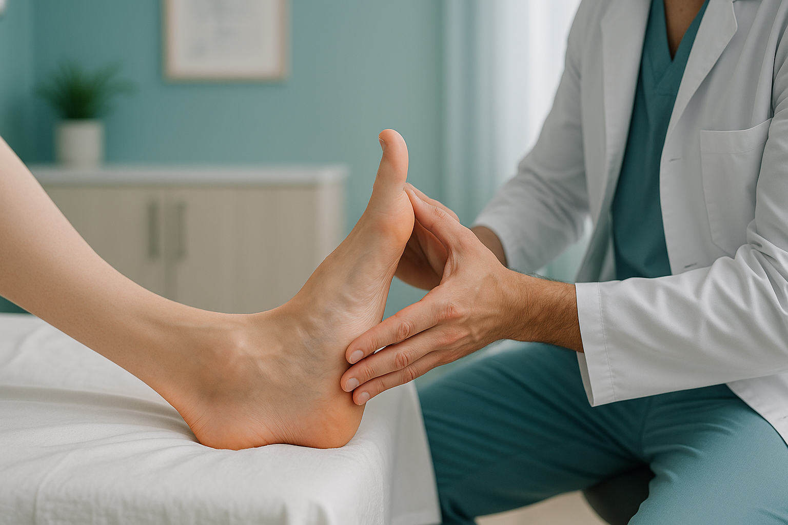
Pes cavus is the medical term for a foot with an unusually high arch. In most feet, the sole makes broad contact with the ground. A cavus foot touches down mainly at the heel and the ball, with little or no contact through the midfoot. This makes the foot rigid and less flexible. It does not absorb shock well when you walk or run.
You can often spot it yourself. Stand on a wet surface and look at your footprint. A high-arched foot leaves only a thin strip along the outer edge, or no strip at all. In more marked cases, the foot may look almost "C-shaped" from the side.
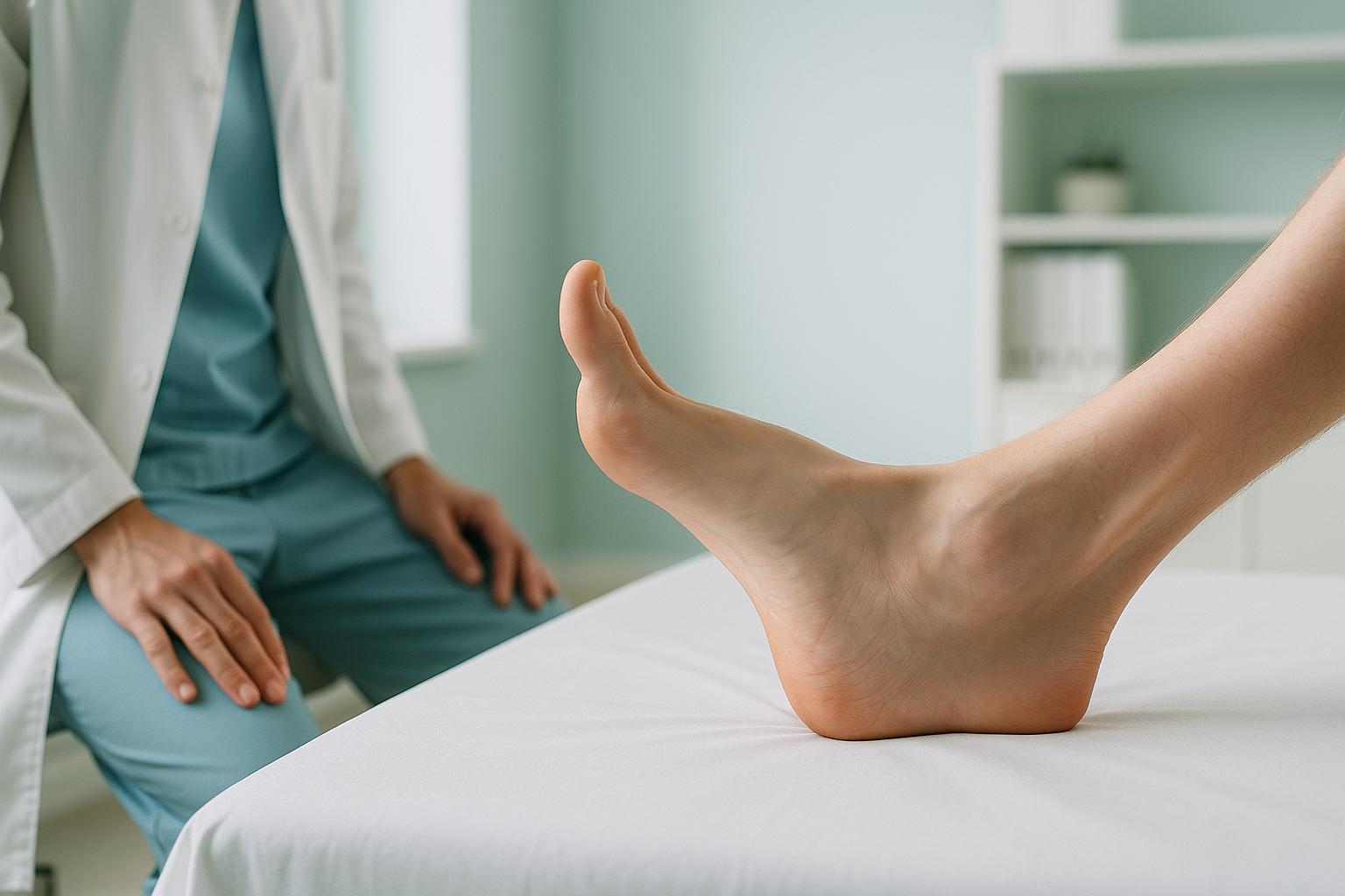
High arches are the opposite of flat feet. A flat foot overpronates (rolls inward). A cavus foot tends to supinate (roll outward). Both can cause problems, but in different ways.
What are the signs that high arches are causing problems?
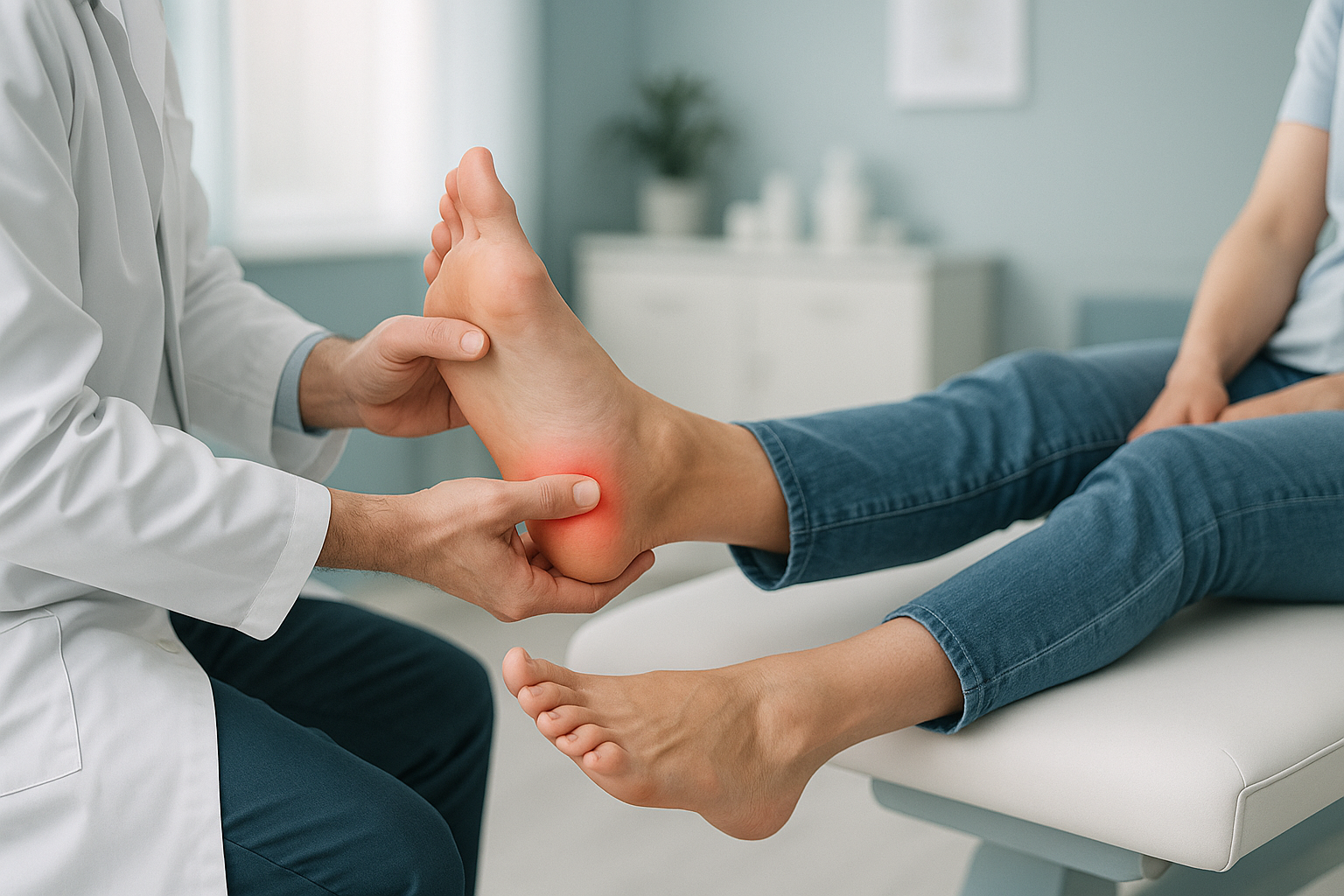
Many people with high arches have no symptoms at all. Problems tend to start when the foot cannot cope with the load you put through it. This is most likely during exercise or long periods on your feet.
If pain on the outer ankle keeps coming back, it is worth reading about peroneal tendonitis on the outer ankle. It is closely linked to how a cavus foot spreads load.
What causes high arches?
A high arch develops when the muscles and tendons that shape the foot are out of balance. This can happen for several reasons.
It helps to understand muscle imbalance and loading. When some muscles in the foot and lower leg overpower others, the arch height and the position of the heel shift. Over time, this can become a fixed change in structure.
Can high arches cause knee, hip or back pain?
Yes, they can. Your body works as a connected chain from your feet to your head. When the foot supinates, the shin turns outward slightly and the knee moves along a different path. The hip and pelvis may tilt to make up for it. Pain often appears well above the foot. If nobody looks at the foot, the real cause can be missed.
This is a common story. Someone has ongoing pain on the outer knee or stiffness in the lower back. Tests focus only on the painful area. The rigid, high-arched foot that started the chain reaction is never examined properly. Understanding how leg alignment is assessed from the ground up can make a real difference.
How is a high arch assessed with standing imaging?
A standard X-ray or MRI is usually taken while you lie down. When you lie flat, gravity no longer loads your foot and your muscles relax. Your arch may look less high than it really is. So the image does not show how your foot truly behaves when you stand and walk.
Standing EOS imaging at ScanAlign captures your feet, legs, hips and spine in one upright image while you carry your full body weight. This shows the real arch height, the angle of the heel (hindfoot varus) and how the rest of the skeleton lines up above. It uses about 90% less radiation than a standard X-ray, around ten times less.
What it shows: True arch height under load, heel tilt, forefoot position, and full alignment from the legs to the spine in one image.
Why it matters: You see how your body really adjusts when it carries weight. This is the information clinicians need to plan orthotics, physiotherapy or surgery accurately.
What it shows: Bone structure and joint spaces without gravity. The arch often looks lower than it really is.
What it misses: Alignment changes that depend on load. The knock-on effects through the knee, hip and spine cannot be seen because the muscles are relaxed.
When MRI is the better tool: EOS shows bones, joints and alignment very clearly. If your clinician suspects soft-tissue damage, nerve compression or a torn tendon, MRI is usually the right choice. The two scans answer different questions and often work well together.
What helps manage pes cavus?
Treatment depends on how severe it is, the underlying cause and your symptoms. ScanAlign is a diagnostic imaging clinic, so we do not provide treatment ourselves. But accurate imaging is the base of any good plan. Common approaches that clinicians may recommend include:
- Cushioned, supportive shoes. Shoes with extra cushioning under the heel and ball help make up for the reduced shock absorption of a rigid, high-arched foot.
- Custom orthotics. Insoles made from your own alignment data can spread pressure more evenly across the sole.
- Physiotherapy. Strengthening the peroneal muscles on the outer lower leg and stretching the plantar fascia and calf can improve stability.
- Ankle bracing. For repeated sprains, a lightweight brace can add support during activity.
- Surgical assessment. In more severe or worsening cases, an orthopaedic surgeon may consider operations to rebalance the foot. Precise standing imaging is essential for planning surgery.
Before you start treatment, or change the one you are on, it is worth getting a detailed diagnosis. Knowing whether the cause sits in the pelvis, the legs or the spine changes what will actually help.
If you have high arches and foot, ankle or leg pain with no clear reason, a standing alignment scan can show what is happening under real load.
Book a free video consultationFrequently asked questions about high arches
Yes. Pes cavus is simply the medical term for high arches. It describes a foot where the arch is higher than the usual range, which puts pressure on the heel and the ball of the foot.
They can. A rigid, supinating foot changes how load travels through the ankle, knee and hip. The body adjusting at any level can cause pain well above the foot itself.
No. ScanAlign is a private, self-pay clinic. You can refer yourself directly through a free video consultation. You do not need a letter from your doctor.
An EOS scan uses about 90% less radiation than a standard X-ray, around ten times less. This makes it one of the lowest-dose options for imaging the skeleton.
It depends on the cause. If high arches are linked to a nerve condition, they may slowly get worse. If the cause is inherited or structural with no underlying disease, the arch shape tends to stay stable, though symptoms can change with how active you are.
The Coleman block test is a clinical examination. You stand with the outer edge of your foot on a small block, and the first metatarsal drops off the edge. It helps a clinician work out whether the heel tilt comes from the forefoot or the hindfoot. This guides treatment decisions.
Orthotics do not change the bone structure. But they can spread pressure, cushion areas under high load and improve stability. Many people with pes cavus find that well-fitted orthotics reduce pain significantly.
When you lie down, gravity is removed and your muscles relax. The arch may flatten slightly and hide how severe it really is. A standing image captures the foot under real body weight. It shows the actual arch height, the heel angle and the alignment of the whole leg above.
The price for patients travelling from abroad is confirmed during the free video consultation, which is how you arrange a foot and leg alignment scan.
If you develop sudden numbness, weakness in the foot or leg, or any loss of bladder or bowel control, get urgent medical help. Contact your doctor, call your local emergency number or go to the nearest emergency department. These symptoms need prompt assessment, not an imaging booking.
See your alignment standing up
A free video consultation lets you talk about your symptoms and find out whether a standing alignment scan in London could help.
Book a free video consultationSources
- NHS. "Foot pain." nhs.uk. Accessed 2024. Overview of common foot conditions including high arches.
- Burns J, Crosbie J, Hunt A, Ouvrier R. "The effect of pes cavus on foot pain and disability." Clinical Biomechanics. 2005;20(9):877-882.
- American Academy of Orthopaedic Surgeons (AAOS). "Cavus Foot." OrthoInfo. Patient information on diagnosis and management of pes cavus.
Written by Abbas Dhami (Specialist Diagnostic Radiographer)
This page is general information and not a diagnosis.
ScanAlign is the trading name of CoreMed Solutions Ltd and operates under The Harley Street Hospital's CQC licence.