
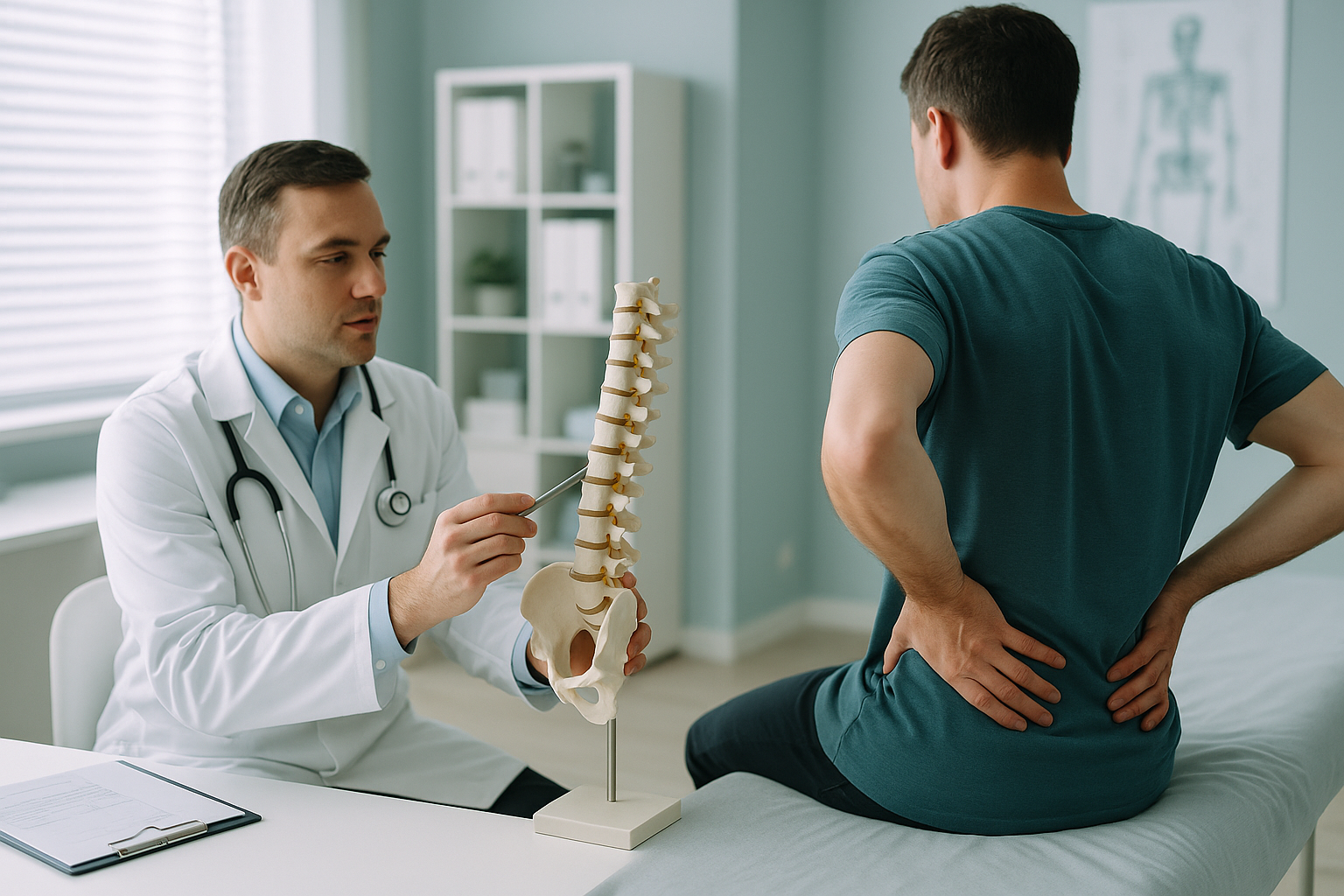
Growth Plate Injury: What Causes It, and Can It Lead to Deformity?
A growth plate injury happens when the soft, developing cartilage near the end of a child's bone is damaged. This is usually caused by a fracture. Most heal well. But if the growth plate is permanently disrupted, the bone may grow unevenly. This causes angular deformity or a leg length discrepancy. Long-term monitoring is what catches these problems early.
Key points
- Growth plates (physes) are the weakest part of a growing skeleton. They are the part most likely to fracture.
- Most growth plate fractures heal without lasting problems, but a small proportion lead to physeal arrest.
- Physeal arrest can cause one limb to grow shorter, or at an angle, compared to the other.
- Regular standing imaging over the years of growth that remain is the only reliable way to spot these changes early.
- Low-dose EOS imaging is well suited to repeated follow-up because it uses about 90% less radiation than a standard X-ray.
What is a growth plate, and why does it matter?
A growth plate, also called a physis, is a layer of cartilage near each end of a child's long bones. It is the area where new bone forms. This lets the limb get longer as the child grows. The physis is made of softer cartilage, not solid bone. So it is structurally weaker than the ligaments and bone around it. That makes it the part most likely to give way under force.
Once a child finishes growing, the growth plates harden into solid bone and are no longer at risk. Until then, any injury to a physis could disrupt how that bone carries on developing.
What are the long-term signs of growth plate damage?
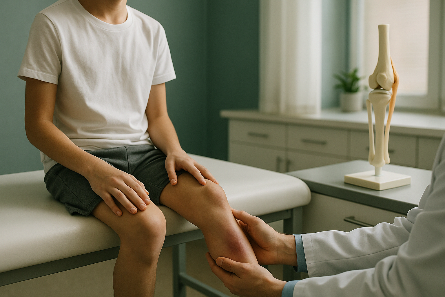
The fracture itself is usually treated at the time of the injury. The real concern is what happens over the months and years after. If part or all of the growth plate stops working, the bone on that side may grow more slowly. Or it may grow at an angle compared to the healthy side. The signs are often subtle at first. They become clearer as the child grows taller.
Clothes may hang unevenly, or one shoe may wear differently. This can point to a leg length discrepancy that is developing.
The limb may start to bow inwards or outwards as the damaged side of the growth plate falls behind.
A change in bone shape can change how a joint moves. This sometimes causes discomfort during activity.
What causes growth plate injuries and physeal arrest?
Most growth plate fractures happen during sport, play or accidental falls. How serious they are depends on how the fracture runs through the physis. Orthopaedic surgeons classify these injuries using the Salter-Harris system. It groups them into types based on the fracture pattern. Higher types carry a greater risk of long-term growth disturbance.
The most common cause. A sudden force, twist or impact can crack the physis.
Salter-Harris types III, IV and V involve the joint surface or crush the physis. This raises the risk of arrest.
A bony bridge forms across the damaged physis. It partly or fully stops growth on that side.
Not every growth plate fracture leads to physeal arrest. Many heal completely, and the child's growth carries on normally. The challenge is that arrest can develop silently over many months. That is why ongoing follow-up imaging is so important.
How is a growth plate injury monitored over time?
Monitoring means imaging both limbs from time to time. This lets the treating team compare growth on the injured side with the healthy side. The child's orthopaedic surgeon will usually ask for scans at set intervals until the growth plates have fully closed. This can mean years of follow-up. This is especially true if the injury happened when the child was young and still has a lot of growing to do.
The key measurements at each follow-up are limb length and joint alignment. Even small differences, when spotted early, can guide the surgeon's decisions. If a significant difference or angular change is developing, options such as guided growth or, in some cases, corrective surgery can be considered at the right time. Waiting until the problem is obvious can make it harder to treat.
When is MRI more suitable? EOS shows bones, joints and alignment clearly. For soft-tissue detail, such as assessing a bony bridge within the growth plate itself, MRI is usually the better tool. Your child's specialist will advise which imaging is needed at each stage.
Why is standing imaging better for growth plate follow-up?
Standing, weight-bearing imaging captures the skeleton as it actually works. When a child lies down for a scan, gravity is removed and muscles relax. Subtle differences in alignment can then be hidden. A standing full-length image of both legs, from hips to ankles, shows the true picture. Any developing tilt, bow or length difference becomes visible in a single exposure.
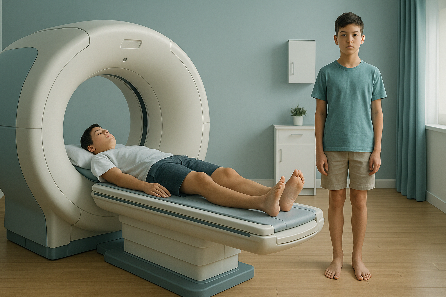
EOS technology is especially well suited to this kind of repeated follow-up. It uses about 90% less radiation than a standard X-ray, around ten times less. A child may need imaging every six to twelve months over several years. So keeping the total radiation as low as possible matters a great deal.
At ScanAlign, low dose standing imaging, each scan produces a full-length, weight-bearing image of both limbs in a single exposure. This gives the orthopaedic team everything they need to track alignment and length accurately. Read more about paediatric imaging at ScanAlign.
Has your child's surgeon recommended follow-up imaging after a growth plate fracture? A low-dose standing EOS scan in London can give the measurements they need with minimal radiation.
Book a free video consultationWhat can be done if a growth plate injury causes deformity?
Monitoring may show that a significant angular deformity or limb length difference is developing. If so, the child's orthopaedic surgeon has several options. The right approach depends on how much growing the child has left and how large the problem is.
- Guided growth. A small plate is placed near a healthy growth plate to slow one side for a time. This lets the limb straighten as it grows. It works best when there is still meaningful growth left.
- Epiphysiodesis. The growth plate on the longer or unaffected leg is closed on purpose at a carefully timed point. This lets the shorter side catch up.
- Corrective surgery. In more severe cases, the bone may be cut and realigned, or lengthened with surgery.
What links all of these is timing. Every one of these decisions depends on accurate, up-to-date measurements of how each limb is growing. That is why regular follow-up imaging matters so much.
Before your child starts a treatment, or changes the one they are on, it is worth getting a detailed diagnosis. Knowing whether the cause sits in the pelvis, the legs or the spine changes what will actually help.
Frequently asked questions about growth plate injuries
No. Most growth plate fractures heal well, and the child's growth carries on normally. Only a proportion, especially the more severe Salter-Harris types, lead to physeal arrest and long-term growth disturbance.
Physeal arrest happens when part or all of a damaged growth plate stops making new bone. A bony bridge forms across the physis. The affected area of the bone stops growing while the healthy side carries on. This can lead to angular deformity or a shorter limb.
Follow-up usually continues until the child's growth plates have fully closed. This can be into the mid to late teenage years. The younger the child at the time of injury, the longer the monitoring is likely to last.
It is possible, but not certain. If part of the growth plate is damaged, the bone may grow at a slight angle over time. Regular imaging picks this up early, so it can be addressed before it becomes a bigger problem.
It is a system doctors use to describe how a fracture runs through the growth plate. There are five main types. Types I and II are the most common and generally carry a lower risk of growth problems. Types III, IV and V involve the joint surface or crush the physis, and carry a higher risk.
EOS captures a full-length, weight-bearing image of both limbs in a single standing scan. This makes it easy to compare limb lengths and joint angles accurately. Because it uses about 90% less radiation than a standard X-ray, around ten times less, it is well suited to repeated scans over several years of follow-up.
No referral from your doctor is needed. You can book a free video consultation directly. Many families bring a request from their child's orthopaedic surgeon, but it is not required.
Yes. EOS uses about 90% less radiation than a standard X-ray, around ten times less. This makes it an especially good choice for children who need repeated imaging over time, as the total dose stays very low.
EOS is designed to show bones, joints and alignment. For detailed soft-tissue assessment, including the inner structure of a growth plate, MRI is usually the better tool. Your child's specialist will advise which imaging is needed.
The price for patients travelling from abroad is confirmed during the free video consultation.
Worried about your child's growth after a fracture?
A short, free video consultation with our team can help you understand whether a low-dose standing scan in London is the right next step.
Book a free video consultationWritten by Abbas Dhami (Specialist Diagnostic Radiographer)
Sources
- Salter, R.B. and Harris, W.R. (1963). Injuries involving the epiphyseal plate. The Journal of Bone and Joint Surgery (American), 45(3), pp.587-622.
- British Orthopaedic Association. Standards for Trauma and Orthopaedics (BOAST): Children's fractures. Available at: boa.ac.uk
- Deschênes, S. et al. (2010). Diagnostic imaging of spinal deformities: reducing patients' radiation dose with a new slot-scanning X-ray imager. Spine, 35(9), pp.989-994.
This page is general information and not a diagnosis.
ScanAlign is the trading name of CoreMed Solutions Ltd and operates under The Harley Street Hospital's CQC licence.