
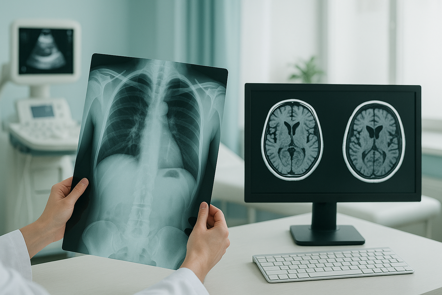
X-ray, Ultrasound, CT Scan or MRI: What Is the Difference?
An X-ray uses a small dose of radiation to show bones. Ultrasound uses sound waves. It works best for soft tissue close to the surface. A CT scan combines many X-ray images into detailed cross-sections. MRI uses a strong magnetic field to give the clearest pictures of soft tissue. Each scan answers a different medical question. The right choice depends on what your clinician needs to see.
Key points
- X-ray and CT use ionising radiation. Ultrasound and MRI do not.
- MRI is generally the best tool for soft tissue such as discs, ligaments and nerves.
- CT gives fast, highly detailed cross-sectional images of bone.
- All four standard scans are done lying down. This can hide alignment problems that only show up when you stand.
- Standing, weight-bearing imaging (such as EOS) captures your skeleton under real load.
You may have been told you need a scan. Or you may be trying to work out which type of imaging will help. Either way, it is easy to feel confused. People mention these names all the time, but nobody explains what each one actually does or why it matters. This guide explains it in plain language.
How does each type of medical scan work?
Each scan uses a different kind of energy to make an image. That energy decides what the scan shows well and what it misses.
A quick beam of ionising radiation passes through the body. Dense parts like bone block more of the beam and show up white on the image. Fast, widely available and low cost.
High-frequency sound waves bounce off tissues and are turned into a live image. No radiation at all. Best for muscles, tendons and fluid-filled structures near the skin surface.
An X-ray tube rotates around you and takes hundreds of images from different angles. A computer builds detailed cross-sectional slices. The radiation dose is higher than a standard X-ray, but the bone detail is superb.
A powerful magnet and radio waves make hydrogen atoms in your tissues give off signals. The result is highly detailed images of soft tissue, including discs, cartilage, ligaments and nerves. No ionising radiation.
What is the difference between CT and MRI, X-ray and ultrasound?
The main difference between a CT scan and MRI is the type of energy used and what it shows. CT uses X-rays and is excellent at showing bone and bleeding. MRI uses magnetism and is excellent at showing soft tissue. The table below compares all four side by side.

| X-ray | Ultrasound | CT | MRI | |
|---|---|---|---|---|
| How it works | Single radiation beam | Sound waves | Rotating X-ray beam | Magnetic field + radio waves |
| Ionising radiation | Low dose | None | Higher dose | None |
| Best for | Bones, fractures, chest | Muscles, tendons, surface soft tissue | Complex fractures, bleeding, organs | Discs, cartilage, ligaments, nerves, brain |
| Bone detail | Good | Limited | Excellent | Moderate |
| Soft-tissue detail | Poor | Good (superficial) | Good | Excellent |
| Contrast dye | Rarely | No | Often | Sometimes |
| Claustrophobia risk | None | None | Mild (short tunnel) | Higher (enclosed, noisy) |
| Patient position | Usually standing or lying | Lying or sitting | Lying | Lying |
Why does it matter whether a scan is done standing or lying down?
It matters because your skeleton behaves differently under load. When you lie down for a CT scan, an MRI or most X-rays, gravity is taken away and your muscles relax. Joints decompress. A disc bulge may shrink. A tilt in your pelvis may flatten out. The image looks reassuring, but it does not reflect how your body actually works when you are upright and walking.
Your body is a connected chain from your feet to your head. A slight shift at one level, for example your pelvis, can make your spine, hip or knee compensate above or below it. You cannot see that compensation on a lying-down scan, because the load has been taken away. This is one reason why many people get scan reports that come back "normal" but still feel pain. The problem is real. The imaging just did not capture it.
Standing, weight-bearing imaging captures your alignment under the same forces your body handles all day. Read more about weight-bearing versus lying imaging and how it applies to back, hip and knee problems.
Not sure which scan would help you? A short video call with our team can make clear whether standing imaging is the right next step, or whether MRI, CT or another scan suits you better.
Book a free video consultationWhich scan should I choose for my problem?
The right scan depends on the medical question, not just the part of the body. Here is a simple guide.
- A fracture is suspected
- You want a quick check of your bones
- You need to see full-body alignment under load
- You are planning joint surgery or having a follow-up after it
- You have disc, cartilage or ligament symptoms
- You have nerve pain, numbness or tingling
- A tendon or muscle tear is suspected
- You need a scan with no radiation at all
Sometimes more than one scan is needed. A standing X-ray or EOS scan can show the alignment picture. An MRI can then show the soft-tissue detail. They answer different questions and often work best together. See how EOS compared with x-ray, CT and MRI fits into the picture.
A note on MRI. EOS and standing X-ray show bones, joints and whole-body alignment. For soft tissue such as discs, nerves, cartilage or ligaments, MRI is usually the better tool. If your clinician suspects a soft-tissue problem, MRI is likely the right place to start. ScanAlign does not offer MRI, but we are always honest about which imaging suits you best.
Before you start a treatment, or change the one you are on, it is worth getting a detailed diagnosis. Knowing whether the cause sits in your pelvis, your legs or your spine changes what will actually help.
Frequently asked questions about types of medical scans
CT uses ionising radiation (X-rays). It is excellent at showing bone detail, bleeding and organ injuries quickly. MRI uses a magnetic field with no radiation. It gives far better images of soft tissue such as discs, ligaments, cartilage and nerves. The right choice depends on what your clinician needs to see.
MRI does not use ionising radiation, so there is no radiation dose. A CT scan does give a radiation dose, though modern scanners keep it as low as reasonably possible. Both are considered safe when used appropriately. Your clinician will recommend whichever one best answers the medical question.
Ultrasound can show the surface of bone, but it cannot see through it. It is not a good tool for fractures or joint alignment. It is best suited to soft tissue near the skin surface, such as tendons, muscles and fluid collections.
At ScanAlign, private standing imaging in London, you do not need a referral from your doctor. You can book a standing EOS scan yourself. For MRI or CT at other clinics, referral rules vary. Check with the clinic directly.
A CT scan gives detailed cross-sectional images. It shows bones in high detail, including complex fractures. It also shows organs, blood vessels and some soft-tissue structures. Contrast dye is often used to highlight blood flow or tumours.
X-rays are fast and widely available. They are the standard first step for suspected fractures, chest problems or basic joint checks. If the X-ray does not explain your symptoms, your doctor may then ask for an MRI to take a closer look at the soft tissue.
EOS is a standing, weight-bearing X-ray system that captures your full body from head to feet in a single image, using about 90% less radiation than a standard X-ray, around ten times less. Because you stand, it shows your real alignment under load. Standard X-rays, CT and MRI are almost always done lying down.
For ScanAlign, the price for patients travelling from abroad is confirmed during the free video consultation. You can find more answers on our frequently asked questions page.
Some metal implants are safe in an MRI scanner and some are not. Older pacemakers, certain aneurysm clips and some cochlear implants are usually not compatible. Your MRI clinic will check you carefully before the scan. If MRI is not suitable, CT or EOS may be an alternative, depending on the medical question.
The best scan depends on your symptoms and what your clinician suspects. If you are unsure, you can check which scan suits you in a free video consultation. Our radiographer can advise whether standing imaging, MRI or another option would help most.
Not sure which scan you need?
Talk to our specialist radiographer in a short, free video call. No referral needed, no obligation.
Book a free video consultationSources
- NHS. "CT scan." nhs.uk, 2023.
- NHS. "MRI scan." nhs.uk, 2023.
- The Royal College of Radiologists. iRefer: Making the best use of clinical radiology, 8th edition.
Written by Abbas Dhami (Specialist Diagnostic Radiographer)
This page is general information and not a diagnosis.
ScanAlign is the trading name of CoreMed Solutions Ltd and operates under The Harley Street Hospital's CQC licence.