

What Does It Mean If a Report Says Your Bone Looks Dense?
If your scan report mentions dense bone, it usually means an area of bone looks thicker or harder than normal. In most cases this is harmless. Often it is a bone island or a sign of normal wear and tear. Now and then it points to something that needs a closer look. So the context matters more than the finding itself.
Key points
- "Dense bone" or "sclerosis" on a report is a description. On its own, it is not a diagnosis.
- The most common cause is everyday wear, especially around the joints and the spine.
- A bone island (enostosis) is a very common and completely harmless finding.
- Context decides whether more imaging is needed. This includes your symptoms, your age and how the area looks over time.
- Standing, weight-bearing imaging can show alignment problems that explain why some bones are under extra stress.
What does "dense bone" or "sclerosis" actually mean on a scan?
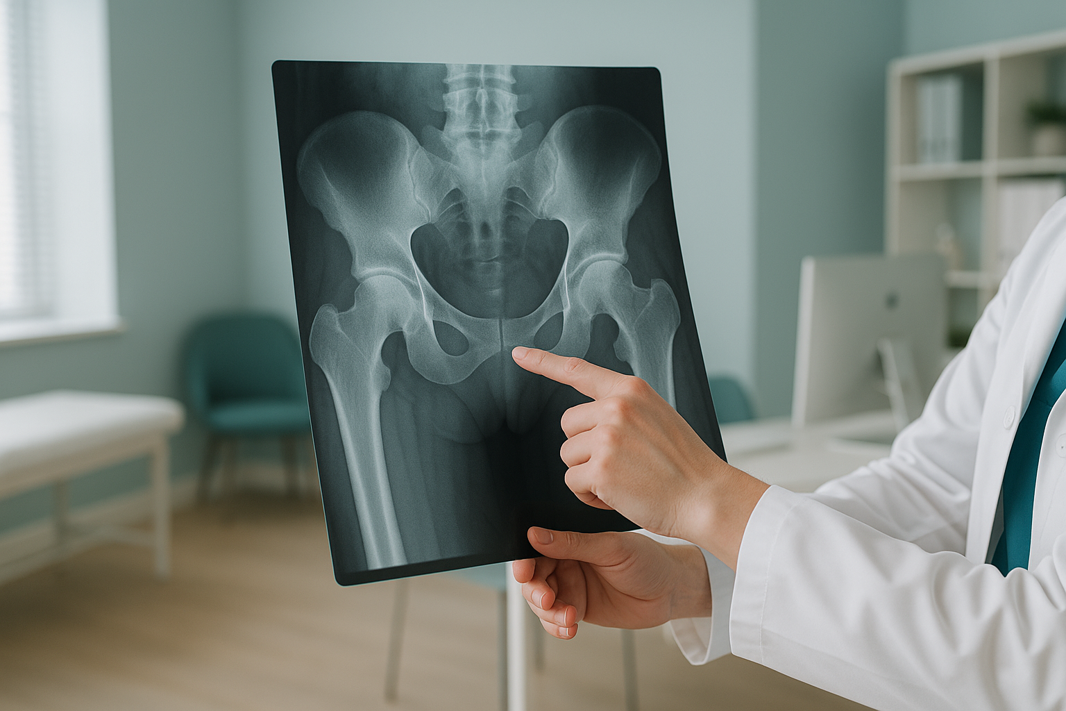
It means the radiologist has seen an area where the bone tissue is thicker, harder or more compact than the bone around it. On an X-ray, dense bone shows up as a brighter white patch. This is because the extra mineral absorbs more of the radiation beam. Radiologists often write "sclerosis" or "sclerotic" in reports. The word simply comes from the Greek word for hard.
This describes how the bone looks. It is not a diagnosis. Think of it like saying a patch of skin is red. The redness itself is not a disease. What matters is why it is red. The same idea applies to a dense area on an X-ray or scan.
What causes a dense area to appear on an X-ray?
Several things can make bone look denser than usual. Most are harmless and need no treatment at all.
A small, compact nugget of dense bone that sits harmlessly inside normal spongy bone. Extremely common. Also called an enostosis.
Everyday wear on the joints or the spine causes the bone under the cartilage to thicken over time. A very common finding in adults.
An old fracture or stress reaction can leave a patch of denser bone behind. This is part of normal healing.
Less common causes
Now and then, a dense area can be linked to conditions that need monitoring or investigation. These include Paget's disease (where bone remodelling becomes disorganised), infection, or, rarely, a growth that needs further assessment. Your doctor or radiologist will look at the shape, size and location of the finding, along with your symptoms. Then they will decide whether more investigation is appropriate.
When should a dense bone finding concern you?
Most of the time, a single dense area on a report is nothing to worry about. But some features lead to further investigation.
A dense area that was not there on an earlier scan, or one that has clearly changed in size, may need a closer look.
If the dense area is exactly where you feel persistent, worsening pain, it is worth discussing with the specialist treating you.
Several dense patches in different bones may suggest a systemic condition that your doctor will want to investigate.
If the finding is a well-defined bone island that has stayed the same on earlier imaging, or a patch of sclerosis near a worn joint, it is almost certainly benign. Comparing it with earlier scans is one of the simplest and most powerful ways to confirm this.
How does standing imaging help explain a dense bone finding?
A dense area near a joint or in the spine often develops because that spot has been carrying more load than it should. Standard imaging is taken while you lie down. Lying down relaxes your muscles and removes the effect of gravity. This can hide the alignment problem that is causing the extra stress in the first place.
A standing, weight-bearing scan captures your skeleton under real load. It shows the whole chain from your feet to your head in a single image. It reveals tilts, shifts or curves that spread force unevenly. If one side of your spine or pelvis is doing more work than the other, standing imaging makes that visible.
ScanAlign standing EOS imaging uses about 90% less radiation than a standard X-ray, around ten times less. That low dose matters if you need comparison scans over time to monitor a finding.
Wondering if a dense bone finding on your report is linked to alignment? A free video consultation, held before you plan any trip to London, can help you understand whether a standing scan is the right next step.
Book a free video consultationWhat should you do after getting a dense bone finding?
Ask your doctor or radiologist to explain the finding in plain words. The two most useful questions are: "Is this likely to be benign?" and "Do we need to compare it with an earlier scan?"
If you have no earlier imaging, or if the finding is in an area where you have ongoing symptoms, more imaging may be recommended. A standing scan can add alignment context that a lying-down image cannot give. It shows whether uneven loading is playing a part in changes in bone density.
When MRI is the better choice: EOS and X-ray show bones, joints and alignment clearly. For soft tissue detail, such as discs, nerves or ligaments, MRI is usually the better tool. The specialist treating you can advise which type of imaging suits your situation.
Before you start treatment, or change the one you are on, it is worth getting a detailed diagnosis. Knowing whether the cause sits in the pelvis, the legs or the spine changes what will actually help.
If you have numbness, weakness, loss of bladder or bowel control, or sudden severe symptoms, please get urgent medical care from your doctor or your nearest emergency department rather than booking a scan.
Frequently asked questions about dense bone findings
No. The vast majority of dense bone findings are benign. Bone islands, degenerative sclerosis and healed injuries are all far more common explanations. A radiologist considers the shape, size and location, and your clinical history, before recommending any further investigation.
A bone island is a small, oval lump of compact bone inside the softer, spongy inner part of a bone. It is completely harmless and does not cause symptoms. It is usually found by chance. It is one of the most common incidental findings on X-rays and CT scans.
"Sclerotic lesion" simply means a patch of bone that is harder than normal. In the spine, the most common cause is degenerative change. This is where years of load and movement cause bone to thicken near worn discs or facet joints. "Lesion" is a general medical term for any area that looks different. On its own, it does not mean anything serious.
Dense bone itself does not usually cause pain. However, the process that created it, such as joint wear or uneven loading, may well be painful. Finding the mechanical cause behind the thickening is often more useful than focusing on the dense area alone.
They look at the shape (smooth and well-defined is reassuring) and the location. They check whether it has changed compared with earlier scans, and whether it matches your symptoms. If everything points to a benign cause, no further action may be needed. If there is doubt, a follow-up scan or more imaging may be suggested.
Not necessarily. Paget's disease has a distinctive pattern on imaging. It often shows enlarged, disorganised bone rather than a small neat patch. A blood test can also help confirm it or rule it out. Most isolated dense findings have nothing to do with Paget's.
A standing scan captures your skeleton under the weight of gravity. It reveals tilts and shifts that disappear when you lie down. If a dense area has developed because one part of your body has been carrying extra load, a standing image can show the alignment problem behind it.
EOS uses about 90% less radiation than a standard X-ray, around ten times less. That very low dose means the extra exposure from an EOS scan is minimal, even if you have had recent imaging. Your radiographer will take your imaging history into account.
No. ScanAlign accepts self-referrals. You can book a free video consultation first to discuss your report. You will find out whether a standing scan would add useful information to your case.
The price for patients travelling from abroad is confirmed during the free video consultation, before you commit to anything.
Have a dense bone finding you want to understand?
A free video consultation with our specialist radiographer can help you work out whether a standing scan in London would add useful context to your report.
Book a free video consultationWritten by Abbas Dhami (Specialist Diagnostic Radiographer)
Sources
- Royal College of Radiologists. iRefer: Making the best use of clinical radiology. RCR, London.
- Greenspan A, Remagen W. Differential Diagnosis of Tumors and Tumor-Like Lesions of Bones and Joints. Lippincott Williams & Wilkins.
- NHS. Paget's disease of bone. nhs.uk (accessed 2024).
This page is general information and not a diagnosis.
ScanAlign is the trading name of CoreMed Solutions Ltd and operates under The Harley Street Hospital's CQC licence.