
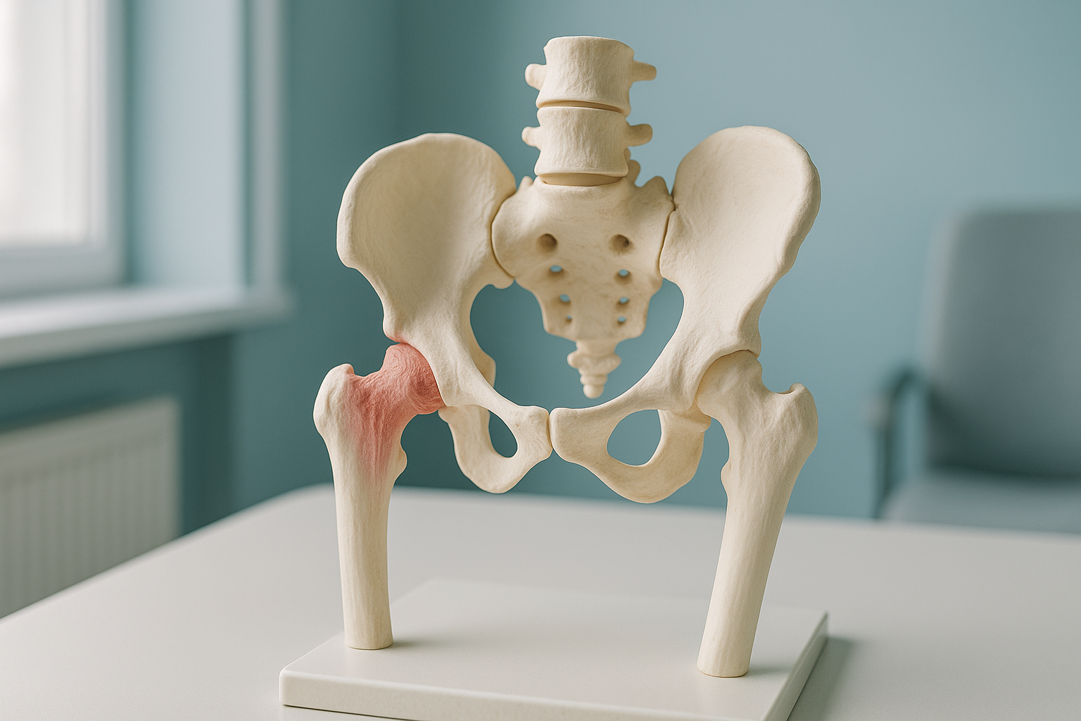
Coxa Vara and Coxa Valga: What Do They Mean for Your Hip?
Coxa vara and coxa valga describe the angle between the neck of your thighbone and its shaft. If this angle is smaller than normal, it is called coxa vara. If it is larger, it is called coxa valga. Both can change how forces travel through your hip. They may contribute to pain, limping or uneven leg length.
Key points
- In a healthy adult hip, the neck-shaft angle is typically between about 125 and 135 degrees.
- Coxa vara (angle too small) tilts the leg inward and can shorten it. Coxa valga (angle too large) does the opposite.
- Both conditions change the leverage of your hip muscles. This can lead to a limp or to extra strain elsewhere as the body compensates.
- Lying-down scans relax the joint and may not show the angle under real weight-bearing load.
- A standing whole-body scan shows how your hip shape works when you are upright and active.
What is the neck-shaft angle of the hip?
The neck-shaft angle is the angle where the neck of your femur (thighbone) meets its main shaft. In a typical adult, this angle is between roughly 125 and 135 degrees. It decides how your hip joint lines up. It also decides how well your muscles can hold you steady when you stand and walk.
When this angle falls below the normal range, it is called coxa vara. The femoral neck points more steeply downward. This tips the leg inward and can sometimes make it functionally shorter.
When the angle is larger than normal, it is called coxa valga. The neck sits flatter, closer to horizontal. This pushes the leg slightly outward and can sometimes make it functionally longer.

Neither term is a disease in itself. Each one describes a variation in shape. But when the variation is significant, it changes how forces pass through your hip joint and the muscles around it. This often causes knock-on problems in other parts of the body.
What are the signs of coxa vara or coxa valga?
The signs depend on how far the angle is from normal and which way it leans. Many people have a slightly unusual angle and feel nothing at all. Larger variations tend to cause more noticeable effects.
The hip muscles lose their mechanical advantage. You may notice a Trendelenburg-type dip, where the pelvis drops on the opposite side as you walk.
A steeper neck (vara) can shorten the leg on that side. A flatter neck (valga) can lengthen it. This may contribute to leg length discrepancy.
Uneven loading through the joint surface can cause a deep ache around the hip or groin, particularly after long walks or standing.
Your body adapts. You may feel pain in your knee, lower back or other hip, rather than in the hip whose structure is different.
What causes coxa vara or coxa valga in adults?
In adults, an abnormal neck-shaft angle usually comes from one of these causes. Sometimes more than one factor plays a part.
The angle may have formed while the bones were growing. It may then carry into adult life without symptoms until wear builds up.
A healed fracture of the femoral neck or shaft can leave the angle permanently changed, especially if the bone healed in a shifted position.
Conditions such as osteomalacia, Paget's disease or avascular necrosis can soften or reshape bone. Over time, this changes the angle.
After hip surgery or an implant, the effective angle can shift. Accurate measurement helps guide planning for any revision surgery.
How is hip shape measured when you are standing?
It is measured with a standing, weight-bearing X-ray that shows the full length of both legs on one image. This shows the true neck-shaft angle under real load. It does not show the relaxed position your bones settle into when you lie on a table.
A standing EOS scan captures both hips, both legs, your pelvis and your spine in one low-dose image. Because you are upright, the image shows how your hip angle behaves under your body weight. Muscles, ligaments and gravity all act naturally. So the measurement reflects how your body actually works.
ScanAlign standing EOS imaging uses about 90% less radiation than a standard X-ray, around ten times less. The full-length view also shows whether one leg is effectively longer or shorter. It shows whether your pelvis or spine is compensating for the hip angle.
A conventional X-ray or CT scan taken while you lie down removes gravity and muscle tension from the picture. The angle measured on a lying-down image can differ from the standing one, because the joint is not carrying load. This may understate the real effect of coxa vara or coxa valga on your alignment.
Lying-down imaging is still useful in some situations, for example straight after a fracture or during surgery. But to understand how your hip works day to day, a weight-bearing image gives a more complete answer.
When is MRI the better choice? EOS shows bones, joints and alignment. If your clinician suspects soft-tissue damage, labral tears or nerve problems around the hip, MRI is usually the better choice. The two types of imaging work well together.
What helps if you have coxa vara or coxa valga?
The right approach depends on how far the angle is from normal, what symptoms you have, and what else is going on in your alignment. ScanAlign is a diagnostic imaging clinic, not a treatment provider. Our role is to give you and your clinician the measurements needed to make clear, informed decisions.
Accurate standing measurements matter because they help your clinician:
- Confirm whether the angle is truly abnormal or within normal variation.
- See whether your pelvis, spine or other hip is compensating.
- Plan any corrective procedure, including the choice of implant, with precise measurements.
- Track changes over time by comparing like-for-like standing images.
Before you start treatment, or change the one you are on, it is worth getting a detailed diagnosis. Knowing whether the cause sits in the pelvis, the legs or the spine changes what will actually help.
If you want to understand your hip shape under real weight-bearing conditions, you can book a standing hip scan in London. Or start with a free video consultation from home to discuss whether it is the right step for you.
Book a free video consultationFrequently asked questions
Coxa vara means the angle between the neck and shaft of your thighbone is smaller than normal. This makes the neck point more steeply downward. This can shorten the leg on that side and change how forces travel through the hip joint.
Coxa valga is the opposite. The neck-shaft angle is larger than normal, so the femoral neck sits closer to horizontal. This can make the leg slightly longer and change the leverage of the muscles around the hip.
In a typical adult, the neck-shaft angle is between about 125 and 135 degrees. It varies from person to person and can change with age or bone conditions.
Yes. A steeper angle (vara) can functionally shorten the leg, while a flatter angle (valga) can lengthen it. This is one of the more common reasons these conditions cause symptoms.
It can. When the angle is significantly reduced, the hip abductor muscles lose mechanical advantage. This can produce a Trendelenburg gait, a distinctive dip of the pelvis on the opposite side with each step.
When you lie down, gravity and relaxed muscles change the position of your bones. A standing image shows the angle under your actual body weight. This is closer to how your hip works in daily activities like walking and standing.
Yes. An EOS scan uses about 90% less radiation than a standard X-ray, around ten times less. It is a quick, non-invasive scan where you simply stand still for a few seconds.
No. ScanAlign accepts self-referrals. You can book directly or start with a free video consultation to discuss whether a scan is the right step for you.
The price for patients travelling from abroad is confirmed during your free video consultation.
Yes. The neck-shaft angle directly affects which implant size and design will restore normal mechanics. Surgeons use accurate angle measurements to choose the right parts and plan the correct positioning.
Understand your hip alignment
A standing scan shows your hip shape under real weight-bearing conditions. Start with a free video consultation from home to find out if travelling to London for it is the right step for you.
Book a free video consultationWritten by Abbas Dhami (Specialist Diagnostic Radiographer)
Sources
- Boese CK, et al. "The femoral neck-shaft angle on plain radiographs: a systematic review." Skeletal Radiology, 2016; 45(1):19-28.
- NHS. "Hip pain in adults." NHS.uk, accessed 2024.
- Deschênes S, et al. "Diagnostic imaging of spinal deformities: reducing patients' radiation dose with a new slot-scanning X-ray imager." Spine, 2010; 35(9):989-994.
This page is general information and not a diagnosis.
ScanAlign is the trading name of CoreMed Solutions Ltd and operates under The Harley Street Hospital's CQC licence.