
What Causes Bow Legs in Adults, and Can They Be Straightened?
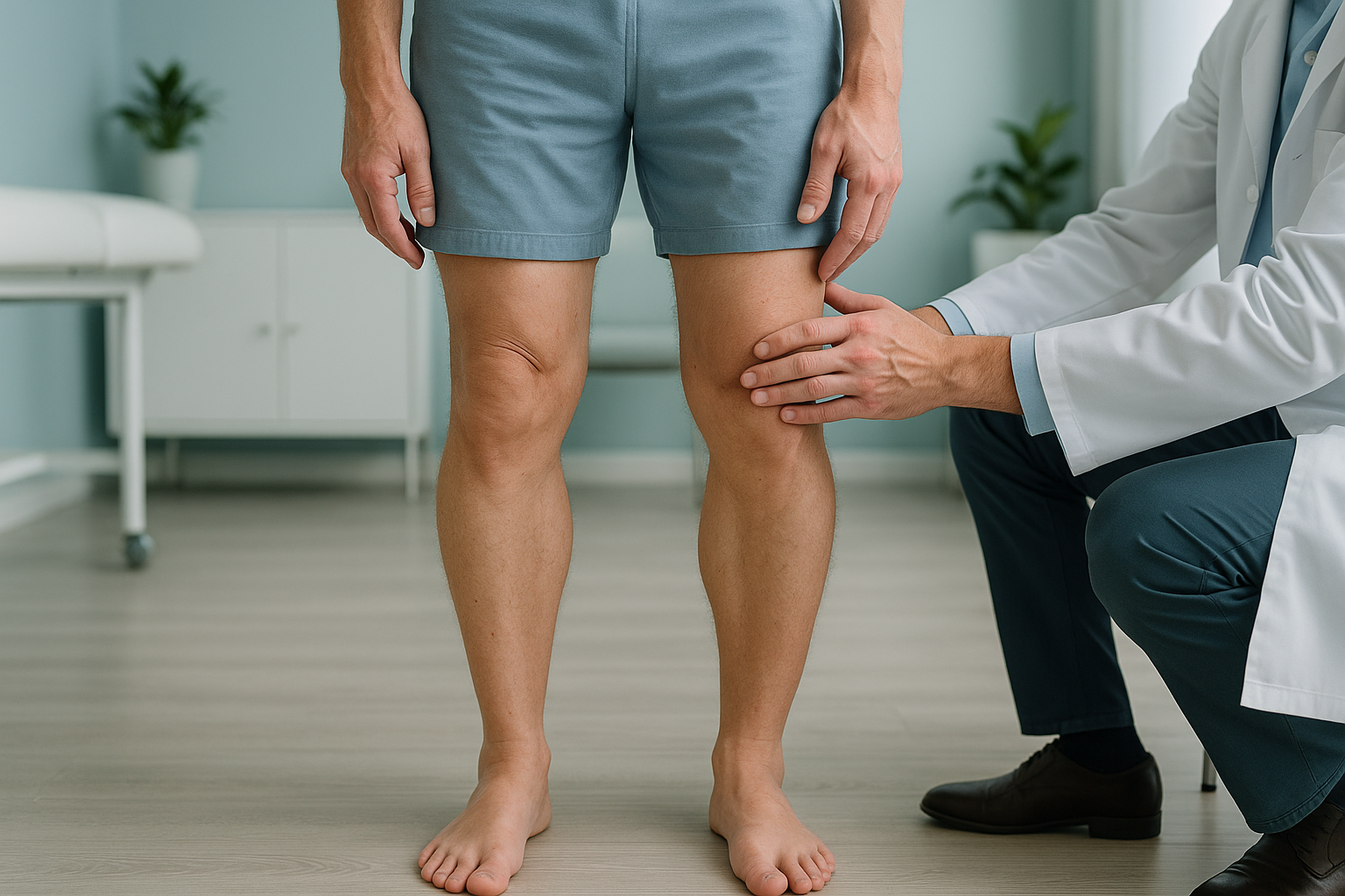
In adults, bow legs (genu varum) are usually caused by slow wear of the cartilage on the inner side of the knee, an old injury, or a childhood condition that was never fully corrected. The outward curve puts extra load through the inner knee. This can speed up arthritis. A standing, weight-bearing scan of the whole leg shows the true degree of misalignment. It helps guide whether a brace, physiotherapy or surgery is right for you.
Key points
- Bow legs in adults are called genu varum. They put uneven load across the knee joint.
- The most common cause in adults is wear-and-tear arthritis on the inner (medial) side of the knee.
- Pain often shows up at the knee, but the root cause can sit higher or lower in the leg alignment chain.
- A full-leg standing x-ray shows alignment that lying-down scans can miss.
- Treatment ranges from exercise and bracing to surgery to realign the leg, depending on how severe it is.
What are bow legs in adults?
Bow legs, known medically as genu varum, means your knees stay apart even when your ankles are together. In a well-aligned leg, a straight line from your hip through the centre of the knee meets roughly the middle of the ankle. If you are bow legged, that line falls on the inner side of the knee instead. This focuses force through the medial compartment of the joint.
A small outward curve can be normal and cause no trouble at all. Problems start when the angle is large enough to overload one side of the knee. The cartilage then wears down faster than it can repair itself. Over time, this uneven load may lead to osteoarthritis, stiffness and pain that limits walking.
How do you know if you are bow legged?
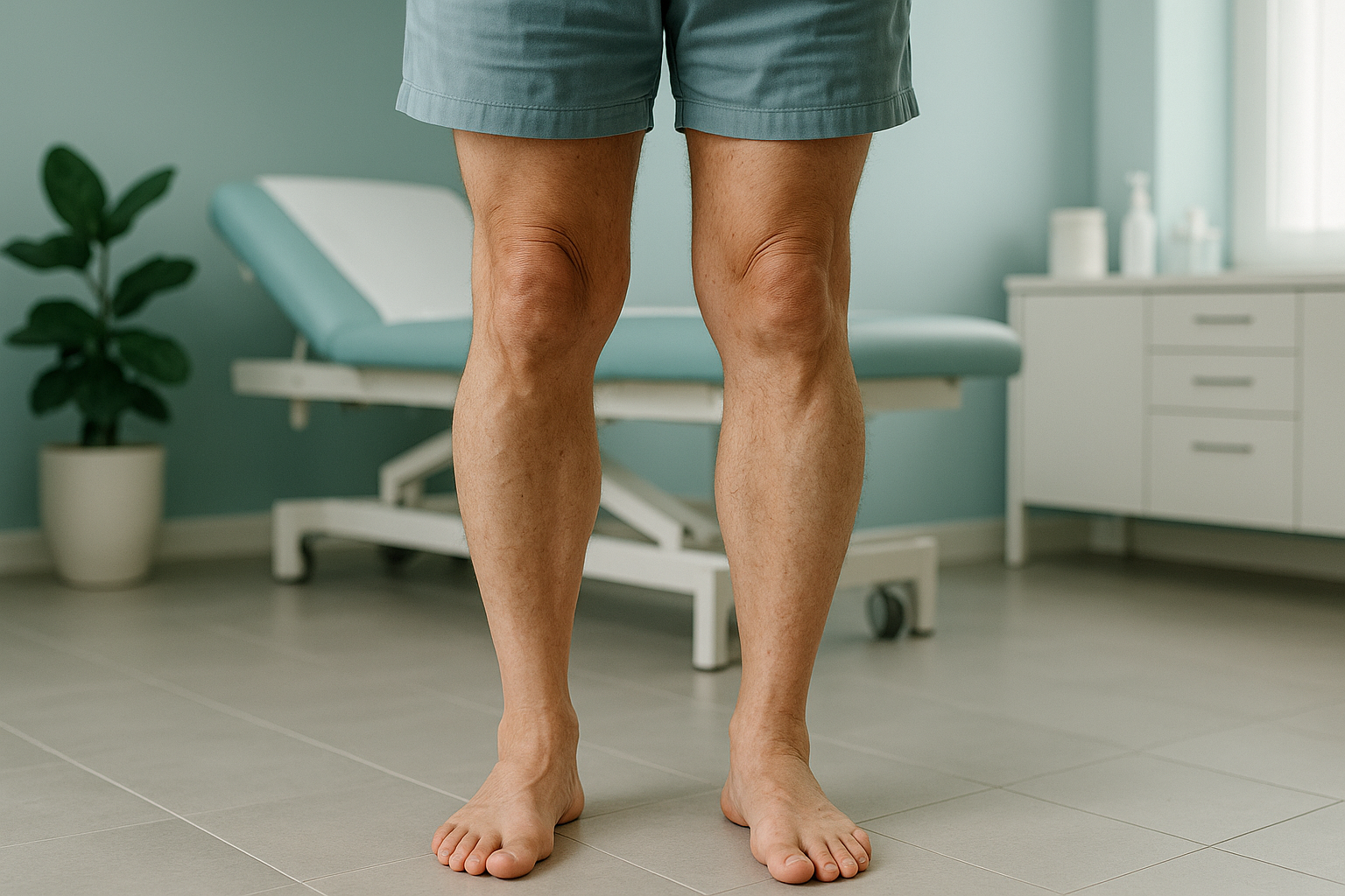
Many people have mild bow legs and never notice. The signs below suggest the alignment may be serious enough to look into further.
When you stand with your ankles touching, a clear space stays between your knees.
Aching or stiffness on the inner side of one or both knees, especially after walking or standing.
The outer edges of your shoe soles wear down faster than the inner edges.
A rolling or wide-based way of walking that feels less steady over longer distances.
What causes bow legs in adults?
In most adults, bow legs develop slowly rather than appearing overnight. The main causes are listed below.
Cartilage on the inner knee wears down. This narrows the joint space and tilts the shin outward.
A broken shinbone or thighbone that healed at a slight angle can leave a lasting varus alignment.
Conditions such as Blount's disease or rickets that were not fully treated in childhood can continue into adulthood.
Paget's disease, vitamin D deficiency or other metabolic bone disorders can soften bone and allow slow bowing.
The body works as a connected chain from feet to head. So a varus tilt at the knee rarely stays on its own. The hip, pelvis and lower back often make up for it to keep you balanced. That is why people with bow legs sometimes feel pain in the hip or lower back rather than in the knee itself.
Why does a standing scan show bow legs more accurately?
A standing, weight-bearing image shows your legs under the same forces they carry every day. Lying down for a standard x-ray or MRI relaxes your muscles and takes away gravity. This partly hides the true curve of the leg. As a result, the misalignment can look smaller than it really is.
A full-leg standing image captures the hip, knee and ankle in one single exposure while you carry your full body weight. The mechanical axis of each leg is measured from the centre of the hip to the centre of the ankle. This shows exactly where force passes through the knee. It is the gold standard for planning any alignment correction.
ScanAlign standing EOS imaging uses about 90% less radiation than a standard x-ray, around ten times less. You simply stand still for a few seconds while the scanner moves from head to foot.
A standard x-ray or CT taken lying down takes away gravity and relaxes soft tissue. The knee may look better aligned than it truly is when you walk. Lying-down images also tend to show only part of the leg. So the overall mechanical axis cannot be measured reliably.
When is MRI the better option? EOS shows bones, joints and alignment clearly. If your consultant suspects soft-tissue damage, a torn meniscus, a ligament injury or nerve involvement, MRI is usually the better tool for those structures. The two scans work well together.
Can bow legs in adults be corrected?
Yes, in many cases. The right approach depends on how much the alignment has shifted and how much it affects your daily life. ScanAlign is a diagnostic imaging clinic, not a treatment provider. Our role is to give you and your clinician a clear, full-leg standing image, so the best way forward can be chosen with confidence.
The options your consultant may discuss usually fall into a few groups.
Strengthening the muscles around the knee and hip to improve support and reduce uneven loading.
A specially designed brace that moves pressure away from the worn inner compartment.
An operation that reshapes the shinbone or thighbone to realign the leg. It is often considered for younger, active patients.
A partial or total knee replacement may be recommended when arthritis is advanced and other options have been used up.
Accurate alignment data from a full-leg scan is especially important before osteotomy or replacement surgery. The surgeon needs to know the exact mechanical axis to plan the correction precisely. Read more about bow legs and knock knees on a standing scan and the difference it makes to planning surgery.
Bow legs are the mirror image of knock knees, where the knees angle inward instead. Both conditions benefit from the same kind of full-leg standing assessment.
Before you start treatment, or change the one you are on, it is worth getting a detailed diagnosis. Knowing whether the cause sits in the pelvis, the legs or the spine changes what will actually help.
Want to understand your alignment before you decide what to do next? You can book a full-leg alignment scan at our London clinic, or start with a free video consultation from home to discuss whether imaging is right for you.
Book a free video consultationFrequently asked questions about bow legs
The most common cause is osteoarthritis wearing down the cartilage on the inner side of the knee. Old fractures, childhood conditions such as Blount's disease, and metabolic bone disorders like vitamin D deficiency can also lead to bowing.
Yes. When the mechanical axis is shifted, extra load passes through the inner knee. This speeds up cartilage wear, which in turn makes the bowing worse. Early assessment can help keep track of any change.
They can. The uneven spread of force through the knee joint often leads to pain, stiffness and swelling on the inner side. Some people also feel discomfort in the hip or lower back as the body makes up for it.
Bow legs (genu varum) curve outward, so the knees stay apart. Knock knees (genu valgum) angle inward, so the knees touch while the ankles stay apart. Both are forms of varus or valgus malalignment and benefit from the same standing full-leg assessment.
Exercise alone cannot change the shape of bone. But targeted strengthening of the hip and thigh muscles can reduce the abnormal load on the knee and ease symptoms. Your physiotherapist or consultant can suggest a programme based on your alignment data.
An osteotomy is an operation where a wedge of bone is removed from or added to the shinbone (or sometimes the thighbone) to realign the leg. It is most often considered for younger, active adults whose arthritis is limited to one compartment.
Standing adds gravity and your body weight. This shows the true degree of bowing under everyday conditions. Lying-down images relax the muscles and can make the alignment look better than it really is.
EOS uses about 90% less radiation than a standard x-ray, around ten times less. It is a quick, comfortable scan where you simply stand still for a few seconds.
No. ScanAlign accepts self-referrals. You can book a free video consultation first, and the clinical team will confirm whether a scan is suitable for you.
The price for patients travelling from abroad is confirmed during the free video consultation.
See your alignment clearly
A full-leg standing scan shows the mechanical axis of each leg under real weight-bearing conditions. Start with a free video consultation from home to find out if imaging is right for you before you plan a trip to London.
Book a free video consultationSources
- Brouwer, G.M. et al. "Association between valgus and varus alignment and the development and progression of radiographic osteoarthritis of the knee." Arthritis & Rheumatism, 2007; 56(4): 1204-1211.
- NHS. "Osteoarthritis: treatment and management." NHS website, accessed 2024.
- Paley, D. "Principles of Deformity Correction." Springer, 2002. Standard orthopaedic reference on limb alignment and osteotomy planning.
Written by Abbas Dhami (Specialist Diagnostic Radiographer)
This page is general information and not a diagnosis.
ScanAlign is the trading name of CoreMed Solutions Ltd and operates under The Harley Street Hospital's CQC licence.