
Why Does a Badly Healed Fracture Change Your Leg Alignment?
Sometimes a broken bone heals in a slightly shifted position. Doctors call this a malunion. Even a small change in angle or length can tilt the mechanical axis of your whole leg. This changes how weight passes through the hip, knee and ankle. It often causes pain, uneven wear and a limp that gets worse over time.
Key points
- Malunion means a fracture healed in an abnormal position. This changes the shape or length of the bone.
- Even a small shift can redirect weight through your leg and cause pain years later.
- Scans taken lying down can miss alignment problems that only appear when you stand and bear weight.
- A standing, full-leg image shows the true mechanical axis from hip to ankle.
- Accurate alignment data helps surgeons decide whether a corrective osteotomy is needed.
What is malunion and how does it happen?
Malunion is the medical term for a fracture that has healed, but in the wrong position. The bone may have joined at an angle, with a twist, or slightly shorter than before. It is not the same as a non-union, where the bone does not heal at all. In malunion, the bone is solid. It is just not quite straight.
This can happen for several reasons. The first fracture may have been complex, with many pieces that were hard to line up perfectly. Sometimes the bone shifts slightly inside a plaster cast or splint while it heals. In other cases, the alignment looked acceptable on early X-rays. But the long-term position turned out to be just enough to matter.
You might not notice a problem at first. The body is very good at adapting. But over months or years, that small change in the shape of the bone redirects the forces passing through your leg with every step you take.
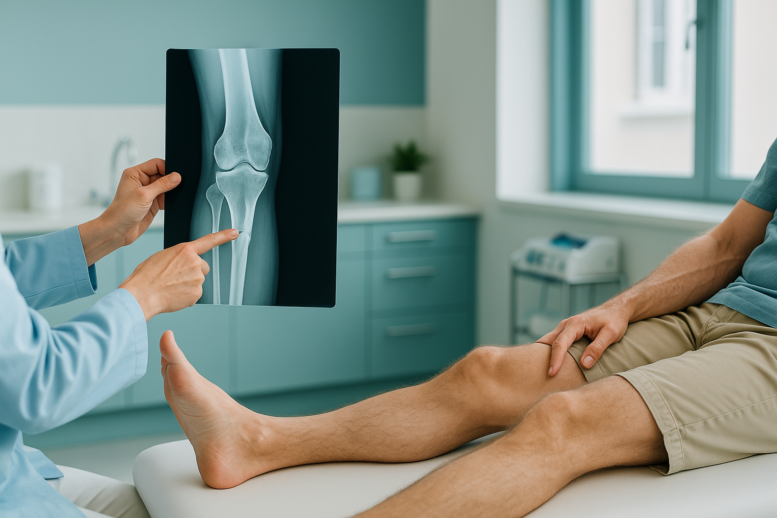
What are the signs of a deformity after a fracture?
The most common sign is persistent or worsening pain in a joint that was not injured in the first place. Because the shape of the bone has changed, the joints above and below take extra stress. These are the patterns people often describe.
Pain in a joint you did not break, especially after walking or standing for a while.
One leg may look slightly bowed, knock-kneed or shorter than the other.
A limp, an uneven stride or the feeling that your body leans to one side when you walk.
Symptoms that were manageable after the fracture but have slowly got worse with time.
If one leg feels shorter after a break, it is worth looking into. A difference in leg length, even a small one, changes how your pelvis sits and how your spine responds. You can read more about leg length discrepancy and the knock-on effects it can have.
How does a malunion affect the rest of your leg?
Your leg works as a connected chain. The hip, knee and ankle are designed to share load in a very specific way, along what is called the mechanical axis. Think of it as an invisible plumb line running from the centre of your hip joint to the centre of your ankle. When the bones are straight, weight passes evenly through the middle of the knee.
A malunion shifts that line. If a shinbone healed with an inward angle, the axis moves to the inner edge of the knee. That side of the joint now carries far more load than it was designed for. Over time, cartilage wears unevenly. Arthritis can develop in a joint that was otherwise healthy.
The body's adjustment does not stop at the knee. Your hip changes its angle. Your pelvis may tilt. Your lower back adjusts further. This is why people with a badly healed broken leg sometimes develop hip or back pain that seems to have nothing to do with the first injury. Understanding lower limb alignment helps make sense of these connected problems.
Why does a standing scan show malunion more clearly than a scan lying down?
A standing, weight-bearing scan shows the true alignment of your leg in real conditions, with gravity pulling and your muscles working. When you lie down for a standard X-ray or CT scan, the muscles relax and the joints decompress. The bones can shift into a position that looks better than it really is. The functional deformity, the one you actually walk on, can be hidden.
An EOS scan captures your whole leg in one standing image, from the hip joint to the ankle, in a single exposure. This means your specialist can see the full mechanical axis and measure exactly where it has shifted. There is no need to join separate images together, which can introduce errors.
EOS also uses about 90% less radiation than a standard X-ray, around ten times less. This matters if you need follow-up scans to track changes or to plan a corrective procedure.
At ScanAlign, standing EOS imaging in London, you stand naturally during the scan. So the image shows how your body actually carries its weight every day.
When MRI is the better choice: EOS shows bones, joints and alignment clearly. If your specialist needs to examine soft tissue, cartilage, ligaments or nerves, MRI is usually the better tool. The two work together. They do not compete.
What can be done about a badly healed fracture?
The first step is getting an accurate picture of what has changed. Without a full-length, weight-bearing image, it is hard to know exactly how much the alignment has shifted and how it is affecting the joints around it.
Once the deformity is clearly measured, your orthopaedic surgeon can decide on the best approach. The options depend on how severe the deformity is and where it is. But they broadly fall into two paths.
- Conservative management. Physiotherapy to strengthen the muscles around the affected joints, orthotics or shoe raises to make up for a length difference, and changes to activity to manage symptoms.
- Corrective osteotomy. A surgical procedure where the bone is carefully re-cut and realigned to restore a more normal axis. It is planned using precise measurements from imaging. This is why accurate data matters so much.
ScanAlign does not provide treatment. It provides the detailed alignment imaging that helps your surgeon or physiotherapist make the right decision. If you want to see exactly where you stand, you can book a full-leg alignment scan and take the results back to your specialist at home.
Before you start a treatment, or change the one you are on, it is worth getting a detailed diagnosis. Knowing whether the cause sits in the pelvis, the legs or the spine changes what will actually help.
Not sure if a standing scan is right for you? A free video consultation lets you talk through your situation with a specialist radiographer from home, before you commit to anything or plan travel to London.
Book a free video consultationCommon questions about malunion and leg alignment
Malunion means the bone has healed, but in an abnormal position. Non-union means the bone has not healed at all, and there is still a gap or movement where it broke. Each needs different management, but malunion is the one that changes alignment.
Yes. Many people feel fine at first because the body adjusts. Over time, the uneven load on nearby joints can cause cartilage wear, pain and stiffness that slowly get worse. It is common for symptoms to appear years or even decades after the fracture.
A change in the angle of the bone shifts the mechanical axis of your leg. Weight that should pass through the centre of the knee is redirected to one side. That side of the joint is overloaded. This can cause pain, swelling and early arthritis, even though the knee itself was never injured.
It can be. If the bone healed with some overlap or collapse where it broke, the leg may end up slightly shorter. A standing full-leg scan is the most reliable way to measure any difference accurately.
It is a planned surgical procedure where the bone is carefully cut and repositioned to restore a more normal alignment. The decision to go ahead depends on detailed measurements from imaging. ScanAlign provides the imaging, not the surgery itself.
No. ScanAlign is a private, self-pay clinic. You can refer yourself directly. Many people book a free video consultation first to discuss whether a scan suits their situation.
An EOS scan uses about 90% less radiation than a standard X-ray, around ten times less. This makes it particularly suitable if you need several images over time to track changes or plan a procedure.
EOS takes a front view and a side view at the same time while you stand. This gives very good information about angular and some rotational deformity. For a complex rotational assessment, a CT scan may also be needed alongside the EOS images.
When you lie down, gravity is no longer loading the leg. Muscles relax and the joints decompress. The alignment on an image taken lying down may not match the alignment you actually walk on. A standing scan shows the true weight-bearing picture.
ScanAlign is a private, self-pay service. For patients travelling from abroad, the price is confirmed during the free video consultation. You can find more details on the FAQs and cost page.
See where your alignment really stands
A free video consultation lets you discuss your situation with a specialist radiographer from wherever you are, with no obligation to book a scan.
Book a free video consultationWritten by Abbas Dhami (Specialist Diagnostic Radiographer)
Sources
- Paley, D. Principles of Deformity Correction. Springer, 2002. A standard reference on limb-alignment assessment and corrective osteotomy planning.
- McKee, M.D. et al. "Malunion of the Long Bones." Journal of the American Academy of Orthopaedic Surgeons, 2016. Overview of malunion classification and management principles.
- UK NHS guidance on broken bones and fracture aftercare. nhs.uk/conditions/broken-leg.
This page is general information and not a diagnosis.
ScanAlign is the trading name of CoreMed Solutions Ltd and operates under The Harley Street Hospital's CQC licence.