
What Is Avascular Necrosis of the Hip, and How Is It Detected?
Avascular necrosis of the hip (also called osteonecrosis of the femoral head) happens when the blood supply to the ball of the hip joint is disrupted. Without blood, bone cells die. The femoral head can then weaken and collapse. MRI is the first-line test for finding it early. A standing scan then shows how the damaged hip affects your alignment under load.
Key points
- AVN happens when blood flow to the femoral head is cut off, causing bone to die.
- Common risk factors include long-term steroid use, heavy alcohol intake, injury and certain blood disorders.
- MRI detects AVN earlier than a standard X-ray, so it is usually the first scan.
- A standing whole-body scan shows how the damaged hip shifts your weight-bearing alignment.
- Finding it early matters. It may open the door to joint-preserving options before the hip collapses.
Your hip joint is a ball and socket. The ball, the femoral head, sits at the top of the thigh bone. It fits snugly into the pelvis. It needs a steady blood supply to stay healthy. When that supply is interrupted, bone starts to break down from the inside. This is avascular necrosis.
AVN can progress quietly. Many people feel only a dull ache at first. It is sometimes mistaken for a muscle strain. Without the right scan at the right time, the condition may not be recognised until the bone surface has already started to crumble.
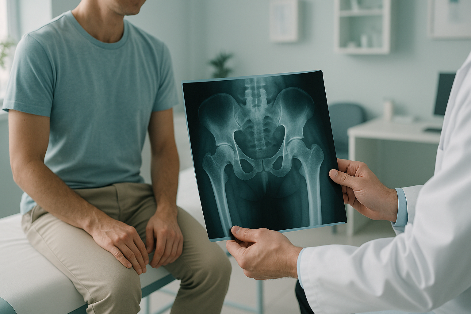
What does avascular necrosis of the hip feel like?
In its earliest stages, AVN may cause no pain at all. As the bone weakens, you typically notice a deep, aching discomfort in the groin or the front of the hip. Pain often gets worse with weight-bearing and eases when you rest.
A dull pain felt deep in the groin or the front of the thigh. It is often hard to pinpoint.
Walking, standing or climbing stairs brings the pain on. Sitting or lying down relieves it.
You may struggle to turn the hip inwards or bring the knee up towards your chest.
As the femoral head weakens, you may start to favour the other leg. This changes the way you walk.
If you develop sudden, severe hip pain, cannot bear weight, or have any numbness or weakness in the leg, seek urgent medical care from your doctor or the nearest emergency department rather than booking a scan.
What causes avascular necrosis of the hip?
Anything that reduces or blocks blood flow to the femoral head can trigger AVN. Sometimes a clear cause is found. In other cases, no single factor is identified.
High-dose corticosteroids taken over weeks or months are one of the most common risk factors.
Regular excessive drinking can damage blood vessels and raise fat levels in the blood. This reduces flow to the bone.
An injury can physically tear or compress the blood vessels that feed the femoral head.
Conditions such as sickle cell disease block small blood vessels. This puts the femoral head at risk.
Other less common triggers include radiation therapy, autoimmune conditions and diving-related decompression illness. When no cause is found, the condition is called idiopathic AVN.
How is avascular necrosis of the hip detected?
MRI is the gold-standard test for early detection. It can show changes in the bone marrow and blood supply before any damage appears on a standard X-ray. If your doctor suspects AVN, an MRI should usually be the first step.
Standard X-rays may look completely normal in the early stages. By the time an X-ray shows a clear problem, the femoral head may already be flattening or collapsing. That is why an early MRI matters so much.
A note on MRI and EOS. EOS is an X-ray-based system. It shows bones, joints and whole-body alignment with about 90% less radiation than a standard X-ray, around ten times less. However, for soft tissue, blood supply changes and early bone marrow damage, MRI is usually the better tool. If AVN is suspected, MRI should come first. EOS then adds a different piece of the picture: how your alignment is affected when you are standing and bearing weight.
Why does standing alignment matter when you have AVN?
Standing alignment matters because the damage AVN causes changes the way your body carries its weight. A weakening femoral head shifts your load onto the other leg. It tilts your pelvis and can change the curves of your spine. These compensations often cause pain in places far from the hip itself.
Lying-down scans relax the muscles and take gravity out of the picture. They can hide the pelvic tilt, the leg-length difference and the spinal shift that appear only when you are upright. A standing whole-body image captures all of this in a single exposure, from your ankles to your spine, under real weight-bearing conditions.
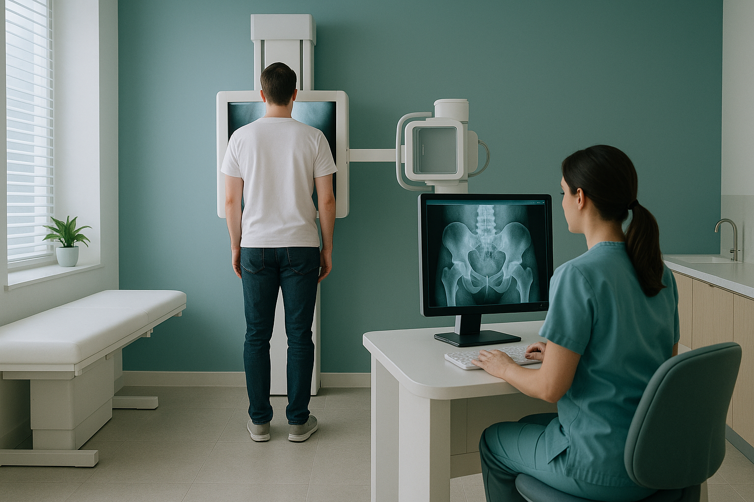
This is what ScanAlign standing EOS imaging provides: a full-length, weight-bearing image that shows how AVN in one hip is affecting the rest of your skeleton. Your consultant can then plan treatment based on the complete mechanical picture, not just the damaged bone on its own.
If you have been diagnosed with AVN and want to understand how it is affecting your alignment, a standing scan at our London clinic can help. No referral is needed.
Book a free video consultationWhat are the general options if AVN is found?
Treatment depends on how far the condition has progressed and how much of the femoral head is affected. ScanAlign is a diagnostic imaging clinic, so we do not provide treatment ourselves. But knowing the general options helps you ask the right questions when you see your specialist.
In early stages, when the bone surface is still intact, joint-preserving procedures such as core decompression may be considered. The aim is to relieve pressure inside the bone and encourage new blood vessels to form. Other approaches include bone grafting or medicines to slow bone loss.
When the femoral head has already begun to collapse or flatten, a hip replacement may become the most reliable option. Standing alignment imaging is especially useful at this point. It helps the surgical team plan the replacement around how you actually bear weight, including any pelvic tilt or leg-length difference that has developed.
If you have hip pain and alignment problems alongside a known AVN diagnosis, understanding both the bone damage and its mechanical effects gives your specialist the fullest possible picture.
Before you start treatment, or change the one you are on, it is worth getting a detailed diagnosis. Knowing whether the cause sits in the pelvis, the legs or the spine changes what will actually help.
Frequently asked questions about avascular necrosis of the hip
Avascular necrosis (also called osteonecrosis) is a condition where the blood supply to the femoral head is disrupted. Without blood, bone cells die. The surface of the hip joint can then weaken and eventually collapse.
Early AVN may cause no symptoms at all. As it progresses, you may feel a dull, deep ache in the groin or the front of the thigh. It gets worse with walking or standing and improves with rest.
Usually not. In the early stages of AVN, X-rays often look completely normal. MRI is far more sensitive. It can detect changes in the bone marrow before structural damage is visible on an X-ray.
MRI is the first-line test for detecting AVN because it shows soft tissue, blood supply changes and early bone marrow damage. EOS is an X-ray-based system that excels at showing how the damage affects your whole-body alignment under weight-bearing. The two work together rather than compete.
MRI is done lying down, which removes gravity. A standing whole-body scan shows how AVN in your hip is affecting your pelvic tilt, leg length and spinal alignment under real conditions. This information is valuable for planning surgery and understanding secondary pain.
Common risk factors include long-term use of corticosteroids, heavy alcohol consumption, hip fractures or dislocations, and blood disorders such as sickle cell disease. In some cases, no clear cause is found.
In early stages, non-surgical or minimally invasive options such as core decompression or medicine may be considered to try to preserve the joint. Your specialist will advise you based on how far the condition has progressed. ScanAlign provides the imaging, not the treatment.
An EOS scan uses about 90% less radiation than a standard X-ray, around ten times less. This makes it suitable for full-body standing images.
No. ScanAlign accepts self-referrals. You can book a free video consultation directly, without a letter from your doctor.
The price for patients travelling from abroad is confirmed during your free video consultation, before you commit to anything.
Understand how AVN is affecting your alignment
A free video consultation lets you discuss your situation with our team from home. You can find out whether a standing scan in London would be useful for you before you travel.
Book a free video consultationWritten by Abbas Dhami (Specialist Diagnostic Radiographer)
Sources
- Mont MA, Hungerford DS. Non-traumatic avascular necrosis of the femoral head. J Bone Joint Surg Am. 1995;77(3):459-474.
- NHS. Avascular necrosis (osteonecrosis). nhs.uk. Available at: https://www.nhs.uk/conditions/avascular-necrosis/
- Karantanas AH. Accuracy and limitations of diagnostic methods for avascular necrosis of the hip. Expert Rev Med Devices. 2013;10(4):545-557.
This page is general information and not a diagnosis.
ScanAlign is the trading name of CoreMed Solutions Ltd and operates under The Harley Street Hospital's CQC licence.