
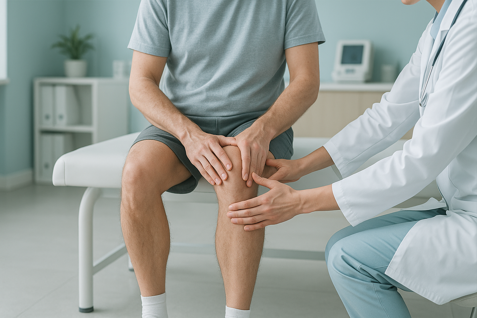
How Does Poor Alignment Increase Your Risk of an ACL Injury?
Poor leg alignment puts extra twisting stress on the anterior cruciate ligament (ACL). This is especially true when the knee collapses inward as you move. Over time, the ligament becomes more likely to tear when you pivot, land or suddenly change direction. A standing whole-body scan can show alignment problems that standard scans miss.
Key takeaways
- Symptoms of an ACL rupture include a pop, sudden swelling, an unstable knee and difficulty putting weight on the leg.
- Alignment problems such as dynamic knee valgus (the knee dropping inward) are a leading risk factor that can be changed.
- Lying-down scans relax the muscles and may hide alignment faults that only appear under load.
- A standing EOS scan shows your full leg alignment from hip to ankle while you bear weight.
- If you think you have a fresh ACL tear, get urgent medical care first, not an alignment scan.
Think you have torn your ACL? Get checked urgently.
If your knee gave way, swelled quickly or feels unstable after an injury, see your doctor or go to the emergency department. You will likely need an MRI to confirm the diagnosis. This page is about understanding and preventing ACL injuries through alignment. It is not about diagnosing a fresh tear.
What are the symptoms of an ACL rupture?
Symptoms of an ACL rupture usually appear straight after the injury. Most people hear or feel a clear pop in the knee. Rapid swelling follows within the first few hours. The knee feels unstable, as if it could buckle at any moment. Putting weight on it becomes painful or impossible.
A popping feeling, or a sound you can hear, at the moment of injury.
The knee often swells a lot within two to four hours.
The knee feels loose or gives way when you try to stand or turn.
Difficulty fully straightening or bending the knee after the injury.
Other ligaments can cause similar symptoms. An MCL injury, for example, also causes swelling and pain, but usually on the inner side of the knee. A careful clinical examination and imaging are needed to tell them apart.
Why do ACL injuries happen?
Most ACL tears happen without contact. They happen when cutting, pivoting, slowing down suddenly or landing from a jump. The ligament fails when the twisting and shearing forces at the knee are more than it can take. Sports with quick changes of direction carry the highest risk, such as football, basketball, netball and skiing.
Several factors raise that risk. Some, like sex or bone shape, you cannot change. Others, like how you move and how well your legs are aligned, you can.
A narrow intercondylar notch, a steep tibial slope or naturally loose joints can make the ACL more vulnerable.
Weak hip abductors and poor landing technique let the knee collapse inward under load.
Leg alignment that favours a valgus (inward-dropping) knee position increases strain on the ACL with every step.
Female athletes have a higher risk of ACL injury than male athletes in the same sports. Research suggests this relates to differences in how they land, the effect of hormones on ligament stiffness, and alignment tendencies.
How does leg alignment affect ACL injury risk?
Your legs work as a chain from hip to ankle. When alignment is off at any point in that chain, the forces at the knee change. The most studied alignment problem linked to ACL tears is dynamic knee valgus. This is when the knee drops inward during weight-bearing activities like squatting, jumping or changing direction.
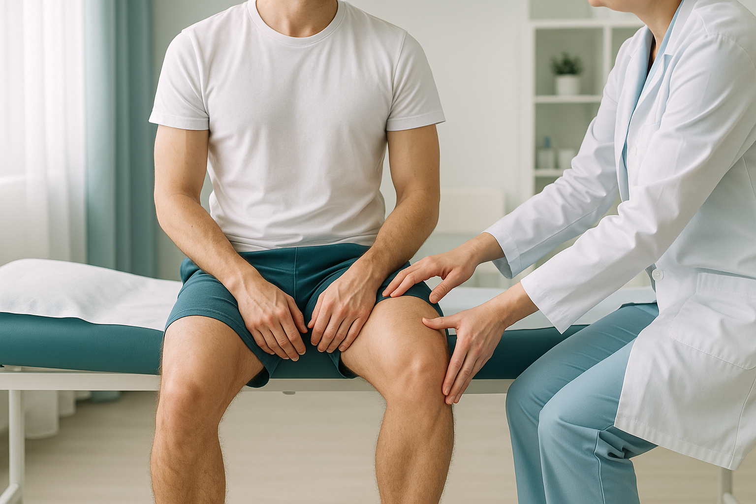
This inward collapse can start at the hip, the knee or even the foot. Weak hip abductor muscles fail to keep the thigh steady. A foot that rolls inward (overpronates) pushes the knee inward from below. If you naturally have knock knees, your starting alignment is already closer to the danger zone. So smaller forces during sport may be enough to overload the ACL.
The key point is that alignment is not just about your knee. It is about the whole chain. A tilt at the pelvis, a rotation at the hip or a stiff ankle can all affect how your knee behaves under stress. This is why looking at the knee alone often misses the bigger picture.
How does a standing scan reveal ACL risk factors?
A standing scan captures your skeleton under gravity. This is the same loaded position your body uses in daily life and sport. It matters because when you lie down for a standard X-ray, CT or MRI, your muscles relax and your joints settle differently. An alignment fault that shows clearly when you stand may simply disappear on a lying-down scan.
An EOS scan images your full legs from hip to ankle while you stand naturally. It shows the true mechanical axis of each leg. It reveals any rotational or angular malalignment, and measures how gravity and your muscles work together. All of this uses about 90% less radiation than a standard X-ray, around ten times less.
Conventional imaging takes pictures while you lie flat. Your muscles switch off, your pelvis levels out, and the ways your body compensates disappear. This can make alignment look normal when it is not. It can miss the very patterns that increase ACL strain during movement.
ScanAlign standing EOS imaging captures a full-length view of both legs in a single low-dose exposure. This gives your surgeon, physiotherapist or sports medicine specialist precise alignment data. They can use it to guide a targeted sports injury prevention programme, or to plan a reconstruction with greater confidence.
When MRI is the right choice: EOS shows bones, joints and alignment under load. For soft-tissue detail, such as confirming an ACL tear, assessing meniscus damage or checking for cartilage injury, MRI is usually the better tool. The two types of imaging work well together.
Can you reduce your risk of an ACL injury through alignment?
Yes. Research consistently shows that neuromuscular training programmes can significantly reduce ACL injury rates, particularly in female athletes. These programmes are designed to improve landing technique, strengthen the hip abductors and correct dynamic knee valgus. Knowing your baseline alignment is a useful starting point. It tells your physiotherapist or strength coach exactly what needs work.
If you have already had an ACL reconstruction and plan to return to sport, an alignment assessment matters just as much. A second ACL injury, to the same knee or the other one, is a well-documented risk. Understanding your structural alignment helps your team make better decisions about when you are ready to return and what ongoing training you need.
Wondering whether your alignment could be putting your knees at risk? A standing scan shows the full picture from hip to ankle.
Book a free video consultationIf you are thinking about an alignment assessment as part of your injury prevention plan, you can book a leg alignment scan without a referral from your doctor.
Before you start treatment, or change the one you are on, it is worth getting a detailed diagnosis. Knowing whether the cause sits in the pelvis, the legs or the spine changes what will actually help.
Frequently asked questions about ACL injuries and alignment
Most people feel or hear a pop, followed by immediate pain and rapid swelling. The knee often feels unstable, as if it might give way. If you have these symptoms after an injury, get urgent medical assessment.
Some people can walk after an ACL tear, but the knee usually feels unstable and hard to trust, especially on uneven ground or when turning. Being able to walk does not rule out a significant tear. Always get it properly checked.
Female athletes are at higher risk because of a mix of factors. These include wider pelvis angles, differences in how they land, the effect of hormones on ligament looseness and a tendency towards greater dynamic knee valgus. Targeted neuromuscular training has been shown to reduce this risk.
Dynamic knee valgus is when the knee drops inward during weight-bearing movements like squatting, jumping or landing. It increases strain on the ACL. It is one of the most important risk factors for ACL tears that can be changed.
A standing EOS scan captures your full legs in one image while you bear weight. This shows your true mechanical alignment. A standard X-ray often covers only one joint and is taken lying down. It may not show alignment faults that appear under load. EOS also uses about 90% less radiation than a standard X-ray, around ten times less.
No. ScanAlign is a private self-pay clinic, and you can book directly without a referral from your doctor. You start with a free video consultation to discuss your concerns and confirm that the scan is right for you.
Yes. The EOS system uses about 90% less radiation than a standard X-ray, around ten times less. It is one of the lowest-dose skeletal imaging methods available.
Yes. Standing alignment data can help your surgeon and physiotherapist assess your recovery and plan when you are ready to return to sport. It can also find any remaining alignment problems that could increase the risk of a second injury.
The price for patients travelling from abroad is confirmed during the free video consultation.
Yes. Poor alignment can increase the risk of other knee ligament injuries, patellofemoral pain, meniscus wear and early cartilage changes. The principle is the same. When forces at the knee are spread unevenly, the structures that absorb those forces are put under greater strain.
Understand your alignment before the next injury
A free video consultation is the first step. We will discuss your concerns, explain what a standing scan would show, and help you decide whether it is right for you.
Book a free video consultationWritten by Abbas Dhami (Specialist Diagnostic Radiographer)
Sources
- Hewett TE, Myer GD, Ford KR, et al. Biomechanical measures of neuromuscular control and valgus loading of the knee predict anterior cruciate ligament injury risk in female athletes. American Journal of Sports Medicine, 2005;33(4):492-501.
- Sugimoto D, Myer GD, Barber Foss KD, Hewett TE. Specific exercise effects of preventive neuromuscular training intervention on anterior cruciate ligament injury risk reduction in young females: meta-analysis and subgroup analysis. British Journal of Sports Medicine, 2015;49(5):282-289.
- NHS. Knee ligament surgery. nhs.uk/conditions/knee-ligament-surgery.
This page is general information and not a diagnosis.
ScanAlign is the trading name of CoreMed Solutions Ltd and operates under The Harley Street Hospital's CQC licence.